


|
|
DOI Prefix 10.20431 |
Information
Journal Policies
Eales Disease in a Young Woman, Case Report
Feria-Anzaldo Estephania MD1,Urzúa De La Luz Pablo MD2,Del Hierro Gutierrez Clarisa Esther MD3,Medina Andrade Abraham Alejandro MD4,Ortiz Ramirez Grecia Yael MD5,Jimenéz-Sierra Juan Manuel MD6,Medina Andrade Luis Angel MD7
2.Glaucoma Department, Association to Prevent Blindness in Mexico, Dr. Luis Sánchez Bulnes Hospital, Vicente García Torres # 46, San Lucas Coyoacán, 04030. Mexico City.
3.Retina Department, Association to Prevent Blindness in Mexico, Dr. Luis Sánchez Bulnes Hospital, Vicente García Torres # 46, San Lucas Coyoacán, 04030. Mexico City.
4.General Surgery Department, General Zone Hospital 1A, Mexican Institute of Social Security, México City.
Copyright :© 2017 Authors. This is an open-access article distributed under the terms of the Creative Commons Attribution License, which permits unrestricted use, distribution, and reproduction in any medium, provided the original author and source are credited.
1. Case Report
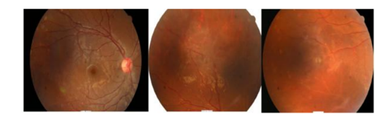
A 17-year-old female patient came to external consultation with a history of sudden loss of vision in the right eye of 3 weeks of evolution. She denies any pathological background, except by previous diagnosis of lichen planus in 2015, currently treated with colchicine and topical steroids.Ophthalmologic exam shows a visual acuity of 20/20 in the right eye and hand motion in the left eye, a normal anterior segment in both eyes, fundus examination in the right eye revealed micro-hemorrhages, bloodless vessels, inferior hypo-pigmented areas, vascular dilation, telangiectasias and phantom vessels (Figure 1); left eye with dense vitreous hemorrhage which did not allow visualization of the posterior pole.
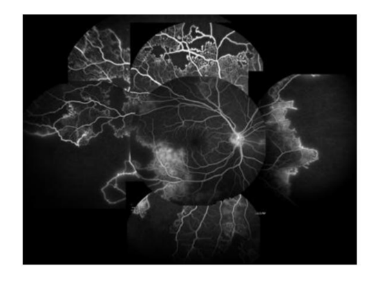
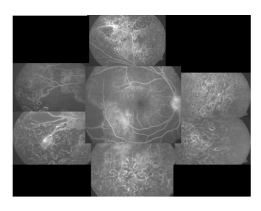
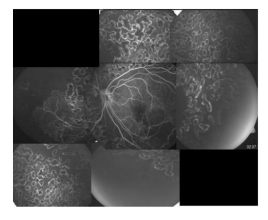
Imaging studies were requested: Fluorescein angiography showed filling defects,telangiectasia vessels, arteriovenous communications and sectorial diffuse hyperfluorescence with an unaltered macula (Figure 2).Given the findings, causes of occlusive vasculitis and vitreus hemorrhage in young patients were assessed. Requested Paraclinical studies were all normal (c ANCA, p ANCA, lupic anticoagulant, anti-cardiolipin, anti beta-glycoprotein, PPD, QuantiFERON TB Gold, VDRL, HLA B-27, rheumatoid factor, C-reactive protein, homocysteine, hemoglobin, hematocrit, blood chemistry and coagulation times). With all the previous information with negative results and normal values, Eales Disease (ED) diagnosis was made by exclusion and a possible association with the previous dermatitis proposed. Patient was treated with vitrectomy and endophotocoagulation on the left eye and pan retinal photocoagulation on the right eye, with a final visual acuity of 20/20 in right eye and 20/40 in left eye.
2. Discussion
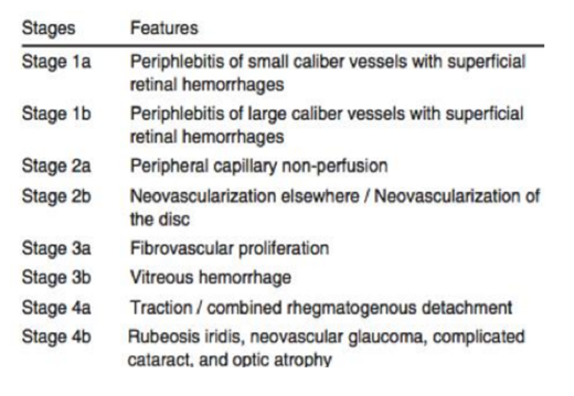
ED is a primary idiopathic obliterative angiopathy of the peripheral retina. It usually affects healthy young adults between 20-40 years old, predominantly males [1] Although unusual in Mexico, Europe and the USA, it is a common cause of visual loss in India and the Middle East [2]. Etiology is unknown, one of the most popular etiologies proposed is related to mycobacterium tuberculosis, though the role of the M. tuberculosis genome in the pathogenesis of ED is yet to be determined [3].It is unilateral at initial stages, but it eventually affects both eyes in 90% of the cases with an asymmetric presentation. It starts first with an inflammatory phase, followed by an ischemic phase and finally a proliferative phase, similar to other proliferative diseases [1]. In our case, we identify our patient in the ischemic stage on the right eye, and the proliferative stage on the left eye. ED usually starts in the peripheral retina and can progress and self limit to the posterior pole [2]. Affected vessels start to obliterate, they stop being functional and appear as fine white lines; these are called phantom vessels. Although in early stages we may observe intraretinal hemorrhages close to the peripheral venules, the most common cause of hemorrhage is the rupture of neovascularization which may produce intraretinal, preretinal or vitreous hemorrhage [4]. Through fundoscopy and fluorescein angiography we may find ischemic perivasculitis, vascular sheathing, retinal neovascularization and recurrent vitreous hemorrhages. Saxena and Kumar proposed a staging system that is useful in classifying and assessing the severity of disease [5] (Table 1).The differential diagnosis is made against other causes of retinal vascular occlusion like sicke cell anemia, systemic lupus erythematosus, Adamantiades-Behçet disease, sarcoidosis, intraocular tuberculsis, etc [6] .Regarding treatment, corticosteroids are still the pillar of therapy in the active perivasculitis stage of ED. Some believe that hypersensitivity to tuberculoproteins take part in the etiology of ED, therefore systemic steroids and anti-tuberculosis treatment is given in patients with retinal periphlebitis with a positive PPD test for a period of 9 months [7].
Macular involvement is uncommon in Eales disease but can occur with central or extensive disease and in the late stages of proliferation8. Periocular steroid injections are used in cases with macular oedema [2].
The most common presentation of ED is a sudden visual loss secondary to vitreous hemorrhage [4]. Initially, the management is conservative and after 3 months, if the hemorrhage has not resolved, vitrectomy with endolaser phtocoagulation is necessary [9].
In the proliferative stages laser photocoagulation it's the treatment of choice, it is unclear which is more effective, either panretinal or segmental photocoagulation [1]. Prophylactic photocoagulation may be used in early stages to prevent secondary complications [10]. Another treatment that can be used in the proliferative stage is anti-VEGF agents, such as bevacizumab. Anti-VEGF agents can be effective in reverting neovascularization and mild hemorrhages [2]. Other studies have found oxidative stress and elevated levels of inflammatory citokines in ED, so current research is aimed at treatments with antioxidant supplementation and immunotherapy aimed to IL-1 and TNF-α [11,12]. The visual outcomes in ED correlate with the clinical stages, early vitrectomy has shown good visual results and prevention of complications, in cases of recurrent vitreous hemorrhage, macular oedema and neovascular glaucoma the final visual acuity is poor [1,9].Our patient presented two uncommon pathologies (lichen planus and Eales Disease), which is why we reviewed previous literature and found only one previous report of bilateral retinal vasculitis in a patient with lichen planus,and by the common autoimmune physiopathology. CD4+ T cell-dependent immunity is thought to play a main role in the pathogenesis of retinal vasculitis, but humoral immunity and immune complex formation may also be important [13]. Cellular immunity plays an important role in retinal vasculitis and lichen planus, and after discarding the other possible causes of vasculitis in the present case we suggest that this is another case related between the dermatitis and ED.
3. Conclusion
Eales disease is a diagnosis of exclusion of occlusive vasculopathies. The treatment of Eales disease depends on the stage of the disease and is not well defined. Observation, pars plana vitrectomy surgery and/or intravitreal injections of anti-VEGF are recommended in cases of vitreous hemorrhage, associated with corticosteroids when retinal vasculitis is present. Laser panretinal photocoagulation is necessary when neovascularization is present. Significant improvement in visual acuity is observed in the majority of stages of Eales disease following treatment. This is the first case of Eales Disease related with lichen planus reported in México and Latin America.
References
- Das T, Pathengay A, Hussain N, Biswas J. Eales’ disease: diagnosis and management. Eye (lond) 2010;24(3):472–82.
- Biswas J, Sharma T, Gopal L, Madhavan HN, Sulochana KN, Ramakrishnan S. Eales disease-an update. Surv Ophthalmol 2002;47:197-214.
- Das T, Biswas J, Kumar A, Namperumalsamy P, Nagpal PN, Patnaik B, et al . Eales' disease. Indian J Ophthalmol 1994;42:3-18
- Kumar D, Saxena RC, Saxena S. Vitreous haemorrhage in Eales' disease. Afro-Asian J Ophthalmol 1995;13:109-12.
- Saxena S, Kumar D: A new staging system for idiopathic retinal periphlebitis. Eur J Ophthalmol 2004,14(3):236–239.
- Agarwal A, Karkhur S, Aggarwal K. Epidemiology and clinical features of inflammatory retinal vascular occlusions: pooled data from two tertiary-referral institutions. Clin Exp Ophthalmol. 2017
- Abu El-Asrar AM, Al-Kharashi SA: Full panretinal photocoagulation and early vitrectomy improve prognosis of retinal vasculitis associated with tuberculoprotein hypersensitivity (Eales’ disease). Br J Ophthalmol 2002, 86: 1248–1251.10.1136/bjo.86.11.1248
- Agrawal S, Agrawal J, Agrawal TP: Intravitreal triamcinolone acetonide in Eales disease. Retina 2006,26(2):227–229. 10.1097/00006982-200602000-00020.
- Asghar A, Niazi JH: Role of vitrectomy in the management of Eales’ disease. Pak J Ophthalmol 2007,23(1):1–5.
- Ishaq M, Niazi MK: Usefulness of laser photocoagulation in managing asymptomatic eyes of Eales disease. J Ayub Med Coll Abbottabad 2002,14(4):22–25.
- Saxena S, Srivastava P, Khanna VK: Antioxidant supplementation improves platelet membrane fluidity in idiopathic retinal periphlebitis (Eales' disease). J Ocul Pharmacol Ther 2010,26(6):623–626. 10.1089/jop.2010.0075
- Saxena S, Pant AB, Khanna VK, Agarwal AK, Singh K, Kumar D, Singh VK: Interleukin-1 and tumor necrosis factor-alpha: novel targets for immunotherapy in Eales disease.Ocul ImmunolInflamm 2009, 17: 201–206. 10.1080/09273940902731015.
- Seyhan Dikci, Oğuzhan Genç, TurGuT Yılmaz, PenPe Gül FıraT. Bilateral retinal vasculitis in a patient with lichen planus. Arq Bras Oftalmol. 2016;79(6):402-3.