


|
|
DOI Prefix 10.20431 |
Information
Journal Policies
Asero-Prevalence of Small Ruminant Brucellosis in Selected
Settlements of Dire Dawa Administrative Council Area,
Eastern Ethiopia
Abu Teshome2, Geremew Haile1*, Lijalem Nigussie3
2.Hawasa University, Collage of veterinary medicine, Hawasa, Ethiopia
3.Jigjiga University, College of Veterinary Medicine, Jigjiga, Ethiopia
Copyright : © 2018 . This is an open access article distributed under the Creative Commons Attribution License, which permits unrestricted use, distribution, and reproduction in any medium, provided the original work is properly cited.
The cross sectional study of small ruminant brucellosis was conducted at 8 selected PAs of Dire Dawa Administrative council from November 2013 to April 2014 to determine the sero-prevalence of small ruminant brucellosis. A total of 424 serum sample are taken; 159 from sheep and 265 from goats and tested for presence of Brucella antibodies against Brucellosis. The sera were first screened for Brucella antibodies using RBPT and positive sera were subjected to the CFT. In this study the overall rate prevalence 2.6% was recorded in small ruminant: 3.02% and 1.9% in goat and in sheep respectively. There was no statically significant difference observed in the prevalence of the disease among different age group, and between sex and species (p>0.05).The prevalence range from o-4.9% were observed in different PAs. Most of the respondent 66.7 % have no awareness about the Zoonotic importance of the disease. 72.2% of flock owners drinks raw milk and 96.3%flock owner handle animals retained fetal membrane with bare hand .In conclusion small ruminant brucellosis were found to be spread in the study area, and the management practice and tradition of using animal’s products exposes the society to Brucella infection.
Brucellosis, Sero-prevalence, Dire Dawa, Small Ruminant,Immunology and Vaccines
1. Introduction
Livestock plays a crucial role in the livelihoods of the majority of Africans. It accounts for 16% of the national and 27-30% of the agricultural GDPs and 13% of the country’s export earnings. The greatest share of this income is from small ruminants [1]. Small ruminants play a big role in supporting the livelihood system of the poorest men and women livestock keepers, especially in the marginalized areas. Sheep and goats are highly adaptable to broad range of environmental conditions. Moreover, low cost of production, requirement of little land and higher prolificacy made them attractive asset for development. Investment in sheep and goats avoid losses due to high inflation rates that are found in unstable economies of many underdeveloped countries like Ethiopia. This is because sheep and goats provide rapid cash turn over [2,3]. This sub-sector receives only very small attention in the poor countries and diseases of small ruminants affect the incomes of smallholder farmers in sub-Saharan Africa by reducing productivity or through loss of the animals[4] .
The small ruminant population of Ethiopia is estimated to be nearly 23.33 million goats and 23.62 million sheep. In the central highlands, Ethiopia, where mixed crop- livestock production system is practiced of small ruminants account for 40% of cash income and 19% of the house hold meat consumption[5].
In spite of the presence of huge small ruminant population, Ethiopia fails to optimally utilize this resource as a sector. This is because of small ruminant production is constrained by the compound effect of diseases, poor feeding, poor management and low genetic endowment. The health and production of animals as well as the wellbeing of humans have been seriously endangered by pathogenic infections. Among these pathogens, different species of brucella are involved in causing brucellosis which is a major disease of domestic livestock and wild animals with serious zoonotic implications in man [6]. One of such disease that hampers the productivity of small ruminants is brucellosis [7,8].
Brucellosis is an acute infectious bacterial disease of domestic, livestock and wild animals which caused by genus Brucella species that has serious zoonotic implications in man; causing huge economic losses to the livestock industry. Cattle, goats, pigs, sheep, horses and dogs play an important role in the transmission of this disease to man. It is also defined as a contagious systemic bacterial disease primarily of ruminants, characterized by inflammation of the genital organs and fetal membranes, abortion, sterility and formation of localized lesions in the lymphatic system and joints [9,10].
The genus Brucella which is causative agent of brucellosis now considered to contain different species: namely, B. abortus, B. melitensis, B. suis, B. ovis, B. canis and B. neotomae. Brucellamelitensis (biovars 1, 2 or 3) is the main causative agent of caprine and ovine brucellosis and it is highly pathogenic for humans causing one of the most serious zoonosis in the world [11]. The disease occurs worldwide, except in those countries where bovine brucellosis (B. abortus) has been eradicated. The disease remains endemic among Mediterranean countries of Europe, Northern and Eastern Africa, Near East countries, India, Central Asia, Mexico and Central and South America. Although B. mellitensis has never been detected in some countries, there are no reliable reports that it has ever been eradicated from small ruminants [12]. Furthermore, brucellosis is also considered as a re-emerging problem in many countries such as Israel, Kuwait, Saudi Arabia, Brazil and Colombia, where there is an increasing incidence of B. mellitensis or B. suisbiovar1 infection in cattle [13].
Brucellosis remains widespread in the livestock populations, and represents a great economic and public health problem in African countries. The epidemiology of the disease in livestock and humans as well as appropriate preventive measures is not well understood, and in particular such information is inadequate in sub-Saharan Africa. In many developing countries, including Ethiopia, a high proportion of the population resides in rural areas where agriculture is the main source of their livelihood. Furthermore, a sizeable proportion engages in livestock production, resulting in a high level of contact with animals and increased risk of Zoonotic infection [14].
In small ruminant, the disease results in significant economic losses due to reproductive impairment caused by abortion, stillbirth or weak lambs and kids, neonatal mortality and, infertility [15]. In humans, brucellosis is often easily misdiagnosed as other febrile syndromes such as malaria and typhoid fever, thereby resulting in mistreatments and underreporting [16]. It is acquired in people through breaks in the skin following direct contact with infected animals’ tissues or blood or their secretions.
Infection may also result from consumption of contaminated unpasteurized milk and milk products. Generally, poor hygiene, prevalence of the disease in animals and practices that expose humans to infected animals or their products influence the occurrence of the disease in humans). Occupational groups at higher risk of infection include cattle producers, veterinarians, animal health personnel, abattoir workers, laboratory personnel and those amongst the general public who are a consumer of animal product. The traditional lifestyle, beliefs and poor knowledge of the disease create favorable conditions for the spread and transmission of Brucellosis. The risks associated with these practices are difficult to control because of a lack of alternatives and simple and/or affordable solutions. The control of brucellosis is likely to be cost effective. Good quantitative information on brucellosis in livestock and the human population is essential for demonstrating the benefits of intervention[17].
Therefore, adequate knowledge of the epidemiology of Brucellosis is of great public health importance, particularly amongst livestock workers and animal product consumers, as this will greatly assist in mapping out strategies for its control. Despite the presence of larger population of small ruminants in different regions of Ethiopia, very limited researches has been done on small ruminant brucellosis, even if it is said to be endemic in the country. The objective of the study is to determine the seroprevalence of small ruminant Brucellosis in and around Dire Dawa.
2. Materials And Methods
The study area, Dire Dawa administrative council, is situated ~ 518 km East of the capital city, Addis Ababa, between 09°28'N to 09°49'N latitude and 41°38'E to 42°19'E longitude. It is situated at an altitude range of 950 to 2250 m.a.s.l., and encompasses an area of 1288.02 km2. The rainfall pattern is bimodal with the highest rainfall in July and August with an average 700 to 900 mm. The monthly mean maximum temperature ranges from 28.1°C in December and January to 34.6°C in May. It is considered the most important area for sheep and goat production. The small ruminant population of the administrative council was estimated to be 227,481 heads (54,600 sheep and 172,881 Goats) [18].
The study animals consisted of 424 traditionally managed small ruminants including 159 sheep and 265 goats above 6 month. The animals were obtained from Adigafelema, Goladey, Dire Dawa, Goro, Bishan Behe, Koriso, Jaldessa and Dujuma peasant associations (PAs). There was no history of vaccination of brucellosis in the study area.
A cross-sectional study was carried out to determine the seroprevalence of small ruminant brucellosis from November 2004 to April 2005. Sera samples of 424 were collected from randomly selected sheep and goats in and around Dire Dawa city. Approximately 7-10ml of blood was drown from jugular vein of apparently healthy adult sheep and goats using plain vacutainer tubes and needles. Individual tubes were identified using numbers and alphabets to indicate their origin, date of collection, species, age, sex. The tubes were left tilt over night at room temperature to allow clotting. The sera was removed from the clot (unretracted blood being centrifuged) by siphoning into sterile test tubes in icebox and stored at -20.
The design adopted for this study was a cross-sectional survey whereby blood samples were taken from randomly selected small ruminants belonging to four peasant associations.
Simultaneously, a questionnaire was administered to small ruminant owning family members. The sample size was determined using the method recommended by [19]for simple random sampling. With an expected prevalence of 50% of small ruminant brucellosis in the selected sites, 0.05% desired absolute precision and 95% level of confidence, the target sample size was calculated to be 384. However, a total of 424 animals (159 sheep and 265 goats) were sampled.
Hence n = (Zx)2 Pexp (1-Pexp) (Thrusfield, 1995) d2 Where: n = the required sample size, Pexp = the expected prevalence rate (50%), Zx = the value of the required confidence interval (1.96) d = desired absolute precision (5%) until serological testing was performed.
The questionnaire was administered only to small ruminant owners in all selected peasant associations by personal interview. The questions were related to the awareness of small ruminant abortions, the consumption of small ruminants’ meat and milk; the practices of handling aborted foetuses and retained foetal membranes.
Prior to blood sampling, data on species, origin, sex and age of the animals were registered. Only sheep and goats older than 6 month were sampled. Blood samples were collected using plain vacutainer tubes and needles directly from the jugular vein and kept overnight to clot at a slanting position at room temperature. Then, the separated serum was carefully collected in a cryovial stored at - 20°C [20] at Dire Dawa Veterinary Diagnostic and Investigation Laboratory until further processing conducted. Furthermore, history of abortion and placenta retention were being recorded.
Rose Bengal plate test (RBPT): The Rose Bengal Plate Test was used as a screening test for the serum samples collected for the presence of brucella agglutinins. The test is conducted as per the procedure recommended by [21,22]. The antigen is obtained from Institute Pourquier, Montpellier, France. This test was carried out at National Veterinary Institution at Debra zeit. The interpretation of the results is done according to the degree of agglutination 0, +, ++ and +++. A score of 0 indicates the absence of agglutination; + indicates barely visible agglutinations; ++ indicates fine agglutination, and +++ indicates coarse clumping. Those samples with no agglutination (0) are recorded as negative while others will record as positive.
Complement fixation test (CFT): All sera which tested positive by the RBPT were retest using CFT for further confirmation. Standard B. abortus antigen for CFT is used to detect the presence of anti-brucella antibodies in the sera. The test antigen is obtained from Addlestone, United Kingdom. The Complement Fixation Test (CFT) were done at National Veterinary Institution (NVI) laboratory at Debra Zeit. Sera with strong reaction, more than 75% fixation of complement (3+) at a dilution of 1: 5 and at least 50% fixation of complement (2%) at a dilution of 1:10 and at dilution of 1:20 will classified as positive [20,23].
The data were analyzed using STATA [24]. Descriptive statistics was employed in determining the prevalence of small ruminant brucellosis and the traditions and practices of small ruminant owners. The logistic regression model was used to identify whether the potential risk factors such as origin of animals, species, sex and age of the small ruminants influenced the sero-prevalence of small ruminant brucellosis. A significant association was said to exist if the Odds ratio (OR) is different from one and the 95% confidence interval of the OR does not include one.
3. Result
Of the 424 animals (159 sheep and 265 goats) the overall sero prevalence of small ruminants were found to be 2.6% (11/424) with the sero prevalence 1.9 % (3/159) and 3.0% (8/265) in sheep and goats, respectively (Table 1).
Table1. Sero prevalence of small ruminant brucellosis in different sex and species
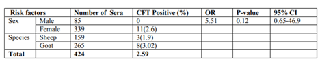
The logistic regression analysis of the risk factors revealed that there is no significant association between some of the risk factors (species age) and the occurrence of brucellosis in small ruminants. On the other hand, sex was found to be important risk factors indicating the significant difference in the occurrence of brucellosis between male and female. In the current study, of the 424 tested small ruminants the entire positives were found to be female animals (3 sheep and 8 goats). To measure the level/strength of association between the brucellosis occurrence and the sex using Odds Ratio cannot be calculated by the employed software (STATA) as the number of positive male animals were zero. But is possible to understand from the Table–2 this is substantial difference between the sexes.
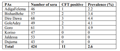
Table2. Sero prevalence of small ruminant Brucellosis in different PAs
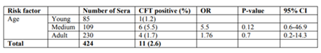
Even though there was no significant difference, a variation in sero-prevalence of Brucella antibody among different age groups was observed from in this study. The sero-prevalence in age groups were (1.2% ), (5.5%) and (1.7%), in young, adult and old age groups, respectively among different age groups, in which the chance of the occurrence of brucellosis was 5.5 times higher in the medium group animals than the young group. There is also higher sero prevalence found in goats (3.0 %) than sheep (1.2%) found in goat and sheep respectively.
4. Discussion
The overall prevalence of small ruminant Brucellosis in this study, based on RBPT, was determined as 3.5% whereas on the basis of CFT, the prevalence was 2.6%.This study demonstrated that the overall individual animal level sero-prevalence of brucellosis in small ruminant was 2.6% (3.0% in goats and 1.9% in sheep). In the present study there is no statically significant difference between sheep and goat. It seems to contradiction with the established facts, in which goat are more susceptible to brucella infection than sheep that could due to the fact sheep do not excrete the organism for long period of time unlike goat. This can mitigate the potential of the spread of the disease among sheep flock [24].
The present investigation recorded a higher sero-prevalence of Brucellosis in goats (3.02%) than sheep (1.9%). These similar results were recorded; 3.2% in goats and 1.6% in sheep in Southern Ethiopia [25], 3.8% in goats and 1.4% in sheep in Eritrea [26] and 4.1% in goats and 1.6% in sheep in East Morocco [27]. Higher sero-prevalence of 16.7% in goats and 14.2% in sheep in Afar [28] and 5.8% in goats and 3.2% in sheep in Afar, Ethiopia [29] .Most breeds of goats are fully susceptible but susceptibility of sheep breeds differs widely [30].
This difference might be due to the differences in flock sizes and proportions of goats and sheep in the herd that is 159 sheep and 265 goats in this present study. In addition, sheep are more resistant than goats and they do not shade the bacteria for long time. Flocks with high numbers of sheep would have low prevalence [31]. Persistent infection of the mammary glands and supramammary lymph nodes is common in goats with constant or intermittent shedding of the organisms in the milk in succeeding lactations, while the self-limiting nature of the disease in sheep, which is seldom accompanied by prolonged excretion of the bacteria[32].
Excretion from the vagina in goats is more copious and prolonged than sheep and lasts for at least 2-3 months. In addition, goats are considered as the principal host of brucella melitensis, whereas, sheep are not significantly infected even when kept in close contact with goats [33]. Infection can vary from a short time occurrence to persistent occurrence for years. In sheep, the course of infection depends upon the dose of infection and after recovery they are resistant to re-infection [30].
In the present study there is no statically significant difference between different sex groups. It seems to contradiction with the established facts, however it is difficult to make firm conclusion as the number of animals is low in study proportion within the factors. The present study showed, CFT positive sera were found only in female animals. The absence of male sero reactor animals in this study could probably be due to the small number of males (N=85) tested as compared to the number of females (n=339). It has also been reported that males are usually resistant than female animals to brucella infection [31,34] have reported that male animals are less susceptible to infection, due to the absence of erythritol. Moreover, it has been reported that the serological response of male animals to brucella infection is limited and testes of infected male animals were usually observed to be non-reactors or showed low antibody titers [35].
A higher prevalence was presently noted in medium age group animals than adult ones. Those at the age of 3 to 5 years (5.50%) were more sero positive than those below three years old (1.8%). However, the variation was statistically non-significant (Table 3) and this variation could be due to the low number of sampled animals in this age group and The placenta is a favored site for replication of the organism large numbers of the organism can be found in chorionic trophoblasts, which contain metabolically active cells capable of producing a variety of hormones and secretory proteins that may stimulate the growth of brucella [31]. Among the selected sites, sero-prevalence of small ruminant brucellosis was highest in sheep and goats sampled from Goro and Dire Dawa; and lowest in that Dujuma, Jeldessa and Koriso although this difference was not found to be statistically significant.
Table3. Sero prevalence of small ruminant brucellosis in different age group
This may due to favoring factors (husbandry, climate, season, lambing): The system of husbandry as well as the environmental conditions greatly influence the spread of infection. Thus lambing/kidding in dark, crowded enclosures is more favorable to spread than lambing/kidding in the open air in a dry environment. The spread of infection between flocks generally follows the movement or gathering of infected animals. The main risk for introducing the disease into a previously non-infected area is by purchase of infected animal (animal movement). Intermingling of flocks may occur under nomadic or semi nomadic conditions of husbandry and also in static village flocks where animals are taken daily for grazing on common pastures are also considered as a factor of different in prevalence’s in different PAs.
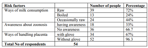
Brucellosis is transmissible from animals to humans through contaminated milk, raw milk products, meat or direct contact with infected animals. During the study questioner survey was collected from different age groups of person (Table 4) on ways handling of fetal membrane, consumption of milk, sharing of night accommodation. From the 54 of the respondents tradition of consuming raw milk 39 (72.2%), sharing night accommodation with their animals 33 (61.1%) and handling of abortion and retained fetal membranes with bare hands 52 (96.3 %), in lack of awareness of the zoonotic nature of the disease 36 (66.7 %) have put the people at high risk of contracting the infection from the animals. It is the fact that the ingestion of raw milk is the main source of infection in human. Milk contaminated with brucellamelitensis is particularly hazardous as it drink in fairly large volume and may contain large number of pathogen organisms [9]. Certain occupations are associated with high risk of infections with brucellosis. These include people who work with farm animals, veterinarians, laboratory staff, and abattoirworkers [9]. The questioner survey also indicates that children and women are more vulnerable to brucella infection as most of activities related with sheep and goat like herding (children 98.1%), milking (children 37.03%, women 96.3%) and milk handling (women 100%) are carried out by them. Some studies show that sero positivity in humans was significantly associated with raw milk ingestion [36,37].
Table4. Questionnaire for risk group people on ways of milk consumption, awareness and ways of handling placenta
5. Conclusion And Recommendation
The result of this study reveals that there is a spread of small ruminant brucellosis in the study area with over all prevalence of 2.6%.Such finding of prevalence in the absence of vaccination against brucellosis indicate occurrence of natural infection. This study also indicate that there is a tradition of consuming raw milk, sharing night accommodation, with animals and handling abortion and retained fetal membrane without wearing protective glove, which in lack of awareness of the disease and its zoonotic nature put the people at high risk of brucella infection. Children and women have been found at high risk of contracting the brucella infection as they carry out most of the activities related with small ruminants. In line with the above conclusion, the following recommendations are forwarded:
• Further epidemiological studies and identification and isolation of the biotype of Brucella responsible for infection in study areas
• A routine vaccination scheme should be practiced in order to reduce its prevalence among animals and subsequently in humans.
• There should be public education about brucellosis in general and its Zoonotic importance.
• Human brucellosis should be considered in the differential diagnosis of certain febrile disease in the study area.
References
- MoARD (Ministry of Agriculture and Rural Development), 2007. Livestock Development Master Plan Study. MOARD, Addis Ababa, Ethiopia.
- Gatenby, R.M. 1991. The Tropical Agriculturalist, Macmillan Education Limited, London and Basingstoke, Pp.1-2.
- Steel, M. 1996. Goats, the Tropical Agriculturalist. Macmillan Education Limited, London and Basingstoke. Pp. 1-2.
- Ibrahim, H., 1999. Diseases of economic importance in small ruminants in sub-Saharan Africa. International Livestock Research Institute, Nairobi, Kenya, Pp: 44.
- Abebe, Z. and Fletcher, I.C. 1993. Small ruminant productivity in Ethiopia mixed farming system. In Proceedings, 4th National Livestock Improvement Conference 13-15 November, IAR, Addis Ababa, Ethiopia.
- Cadmus, S. I. B., Ijagbone, I. F., Oputa, H. E., Adesokan, H. K. and Stack, J.A., 2006. Serological survey of brucellosis in livestock animals and workers in Ibadan, Nigeria. African Journal of Biomedical Research, 9, Pp. 163–168.
- Ademosoum, A.A. 1994. Constraints and prospects for small ruminant research and development in Africa. ILCA, Addis Ababa, Ethiopia, Pp.1-5.
- LMA (Livestock Marketing Authority) 2001. Brief Baseline Information on Ethiopian Livestock Resource Base and Its Trade. Livestock Marketing Authority. Addis Ababa, Ethiopia London and Basingstoke. Pp 1-2. Massachusetts, Pp.196-203.
- WHO, (1986). Joint FAO/WHO Expert Committee on Brucellosis, Technical Report 740, WHO, Geneva, Pp. 86-88.
- CDC. 2005. Brucellosis (Brucellamelitensis, abortus, suis and canis). Available at: www.cdc.gov/ncidod/dbmd/diseaseinfo/ brucellosis_g.htm [Accessed 26 November OIE, 2000. Manual of Standards for Diagnostic Tests and Vaccines. 4thed. Paris, Pp475- 481.
- FAO/OIE/WHO (2002). Animal Health Yearbook, FAO Animal Production and Health Series, FAO, Rome, Italy.
- Cutler, S. J., Whatmore, A. M. and Commander, N. J. (2005). Brucellosis-new aspects of an old disease. Journal of Applied Microbiology, 98 (6), Pp. 1270-1281.
- McDermott, J. J. and Arimi, S. M., 2002. Brucellosis in Sub-Saharan Africa: epidemiology, control and impact. Veterinary Microbiology, 20, Pp. 111-134.
- Xavier, M. N., Costa, E. A., Paixao, T. A. and Santos, R. L., 2009. Genus brucella and clinical manifestations. Ciencia Rural, 39, (7), Pp. 2252-2260
- Bax, H. I., Van Veelen, M. L. and Gyssen, I. C., 2007.Brucellosis, an uncommon and frequently delayed diagnosis. Netherlands Journal of Medicine, 2, Pp. 352–355.
- Swai, E. S. and Schoonman, L., 2009. Human brucellosis: Seroprevalence and risk factors related to high risk occupational groups in Tanga Municipality, Tanzania Zoonoses and Public Health 56, Pp. 183–187
- Council of state administrative Dire Dawa., (2010).
- Thrusfield, M.V., 2005. Veterinary Epidemiology. Z. 3 edition, Blackwell Science, Oxford, Pp.234-238.
- Viriego FJ, Moreno MA, Dominguez L (2000). Risk factors for brucellosis seroprevalence of sheep and goat flocks in Spain. Prev. Vet. Med., 44(3–4): Pp.167–173.
- Alton, G.G., Jones, M.J., and M., Lois, M. and Peitz, D.E. 1975. Serological methods In: Laboratory Techniques in brucellosis.2nded. WHO, Geneva.Pp.64-124.
- Nielsen, K., and Dunkan, J.R. 1990. Animal Brucellosis. CRS Press INC, Pp 173-179.
- OIE, 2004. Manual of Diagnostic Tests and Vaccines for Terrestrial Animals. 5th ed., Office International Epizootics, Paris, Pp 409-438.
- Radostits, O. M., Gay, C. C., Blood, C. D. and Hinchcliff, K. W., 2000. Veterinary Medicine, Textbook of the Disease of Cattle, Sheep, Pigs, Goats and Horses. 9th edition. New York: W.B. Saunders Company Ltd, pp. 867-882.
- Mengistu, M., 2007. Sero epidemiology of brucellosis in small ruminants in southern Ethiopia, M.Sc. thesis, Addis Ababa University, Faculty of Veterinary Medicine, DebreZeit, Ethiopia.
- Omer, M. K., Skjerve. E., Holstad, G., Woldehiwot, Z. and Macmillan, A. Pp., 2000. Prevalence of antibodies to Brucella spp in cattle, sheep, goats, horses and camels in the state of Eritrea; influence of husbandry systems. Epidemiol Infect, 125, Pp. 447 – 453.
- Benkirane, A., 2006. Ovine and caprine brucellosis: World distribution and control and eradication strategies in West Asian and North African region. Small Ruminant Res., 62: 19-25.
- Teshale, S., Muhie, Y., Dagne, A. and Kidanemariam, A., 2006. Seroprevalence of small ruminant brucellosis in selected districts of Afar and Somali pastoral areas of Eastern Ethiopia: the impact of husbandry practice. Revue de` Elevageet Medicine Veterinaire des Pays Tropicaux157, Pp. 557-563.
- Ashenafi, F., S. Teshale, G. Ejeta, R. Fikru and Y. Laikemariam, 2007 Distribution of Brucellosis. Among small ruminants in the pastoral region of Africa, eastern Ethiopia. Rev. Sci. Tech. Off. Inf. Epi., 26: Pp.731-739.
- European Commission (EC), 2001. Brucellosis in sheep and goats (Brucellamelitensis). In: Report of the Scientific Committee on Animal Health and Animal Welfare.
- Radostitis, O.M., Gay, C.C.K.W. and Hinchcliff and P. D. Costable, 2007. Veterinary Medicine .A text book of the disease of the cattle, sheep, pigs, goats, and horse, 10th edition London, Bailliere Tindall, Pp. 992-1003.
- Alton, G.G., (1990). Brucella mellitensis In: “Animal brucellosis”. (Nielsen, K., Duncan, J. R., eds). CRC press. Boston, Pp.383-409.
- Alton, G.G., 1985. Epidemiology of brucella melitensis in sheep and goats: In: brucellamelitensis, a CEC seminar, Eds., J.M. Verger and M. Plommet. Martinus Nijoff, Dordrecht-Bosten.
- Hirsh, D.C and Zee Y.C.1999. Veterinary Microbiology. Blackwell Science, Cambridge, Massachusetts, Pp.196-203.
- Crawford, R., Huber, J.D. and Adams, B.S. 1990. Epidemiology and surveillance. In: Nilsson, K. and Dunkan, J. R. (eds), Animal Brucellosis, CRS Press Inc., Florida, Pp. 131-1486. De. massis, F., Digriolamo, A. and Giovannini, A.2005.Correlation between animal and human brucellosis in Italy during the period of 1997-2002. Jour.Euro.Soci.Micro. and inf. Dis., 11:Pp. 632-636.
- Abraham. 2006. A sero-prevalence study of brucellosis in goat and humans in Chifra woreda, Affar regional state. DVM thesis, faculty of Veterinary Medicine, Addis Abeba University, Debra zeit, Ethiopia.