


|
|
DOI Prefix 10.20431 |
Information
Journal Policies
Stress Fractures of Sacrum
Liimatainen E1, Alonso J3, Lempainen L1,2, Johansson K1, Sarimo J1,2, Frantzen J2, Orava S1,2
2 Hospital NEO and Sports Trauma Research Center, Turku, Finland
3 Sports Medicine Department, Aspetar, Qatar Orthopedics and Sports Medicine Hospital, Doha, Qatar
Copyright : © 2016 Liimatainen E. This is an open access article distributed under the Creative Commons Attribution License, which permits unrestricted use, distribution, and reproduction in any medium, provided the original work is properly cited.
Abstract
Background: Stress fractures of sacrum are rare overuse injuries. They are difficult to diagnose, because usually the symptoms are thought to result from other reasons in the pelvic area.
Purpose: To report a series of sacral stress fractures in athletes and physically active older individuals.
Study design: Case series.
Methods: During the years 1996 – 2009 a total of 40 cases of sacrum stress fractures were treated in Private Hospital Sports Clinic in Turku, Finland and RFEA Clinic, Madrid, Spain. MRI was used for diagnosis in all patients but one.
Results: All athletes were able to start running 2.5 months after the diagnosis and full training was possible after 3.5 months. Initial radiography was negative in all patients.
Conclusion: Early MRI is recommended in endurance runners. Older individuals and amenorrheic females with possibly osteoporotic bones require longer resting period from impact training. Compensative training program is safe and doesn’t interfere the healing of sacrum stress fracture.
Key words: sacrum, stress fracture, insufficiency fracture, athlete, older individuals, sports.
1.Introduction
Stress fractures of in and around the pelvis are uncommon compared to stress fractures and osteopathies in the lower extremities[1,2]. They occur at all ages, both children and elderly[3,4]. Stress fractures of the sacrum can be difficult to diagnose, because symptoms are often thought to relate to other, more common musculoskeletal problems. Athletes’ sacral stress fractures were not seen in a larger extent 25 years ago[5,6]. Sacral stress fractures were first reported as insufficiency fractures in osteoporotic patients[7,8]. In military recruits sacral stress fracture was described to occur following strenuous physical activity [9]. Since then more case reports and small series in athletes and army recruits have been described [2, 4,10,11]. We report a series of sacral stress fractures seen in athletes and physically active older individuals.
2. Patients and Methods
Data was collected retrospectively from patient records. Patients included all treated for sacral stress fractures, excluding malignant etiology. Information for the onset of sympoms and diagnosis as well as patient characteristics were retrieved. During the years 1996-2009 a total of 40 cases of stress fractures of sacrum were seen in patients visiting private Sports Clinic in Turku, Finland and RFEA Clinic, Madrid, Spain. The athletes represented mainly endurance running. There were only one middle distance runner and one walker in the series. Sixteen of the patients were active runners, four middle-aged joggers and three older walking individuals in the series. There was only one senior patient, in addition to them, who was not involved with physical activity at the time of appearance of the symptoms (Table 1).
3. Results
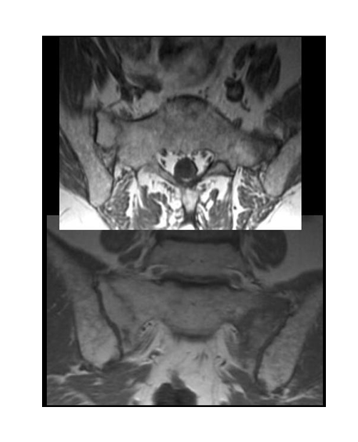
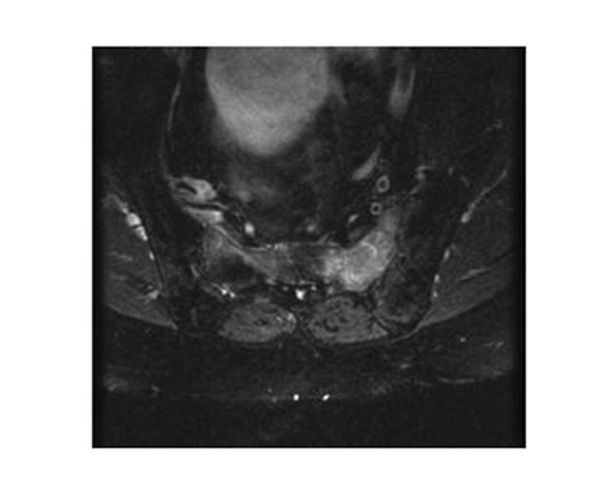
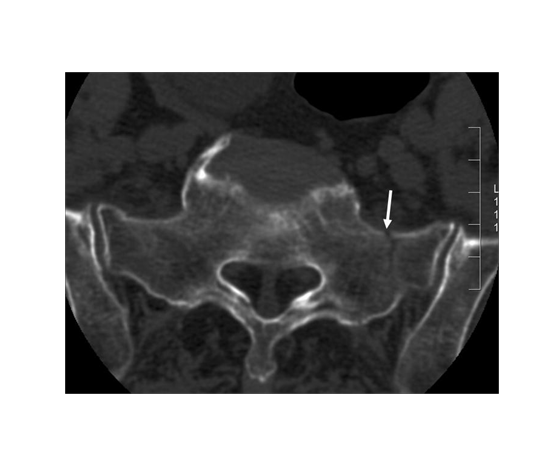
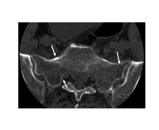
The diagnosis was done in athletes approximately 1.5 months after the onset of the symptoms. In other patients the diagnostic delay was 3.5 months. The initial radiographs did not show the fracture in any of the six examined patients. Isotope scan and CT-examination were done in one of the patients. MRI was used in all other patients, which was diagnostic (Figures one through five).
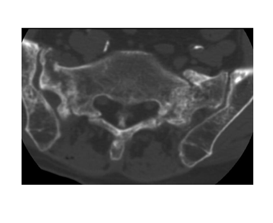
Bilateral stress fracture of sacrum with more bone oedema on the left side
T2 fat supression images on a middle aged runner
MRI was used as follow-up examination in 4 of the patients approximately four months after the diagnosis. At the follow-up there was a clear healing seen at the fracture site. All the patients suffered from lower back and pelvis pain, which was dull and diffuse in the beginning, felt during and after physical exercise. Later the pain continued also in normal daily life (40%) or in the beginning of running preventing it (69%). There were 25 male and 15 female stress fractures in the series. The mean age of the patients was 35.4 years (17-79 years). In active athletes (runners) the mean age was 25.2 years (17-33 years). Two of the patients suffered from sacrum stress fracture first on one side and one year later on the other side of sacrum. Patient characteristics are presented in Table 2.
The treatment started with cessation of running. After an initial 2-3 week rest period swimming and water running among other non-impact type exercises were allowed. Cycling and gym training were added to the athletes compensative training one month later and rowing as well as cross-country skiing was allowed one month later. All athletes were able to start running 2.5 months from the diagnosis and full training was possible one month later. Older patients required two months more before returning to full activity.
4. Discussion
Lower back and sacral pains during and after physical exercise in active young people are at first considered as musculotendinous pain or originating from vertebral facets or discus [2,4,12,13]. If pains become chronic, radiographic examinations are made. The findings may show spondylolytic defects, anomalies, posterior iliac apophysitis, sacroiliac changes or stress fractures of sacrum [2], [14,15]. True stress fractures of sacrum create diffuse pain located at the sacrum, posterior lower back and pelvis area. The pain usually becomes more pronounced with training and may prevent running, which usually has been reported as the most common causative factor[3]. Pain is typically posterior hip and buttock pain in unilateral cases[4,16]. Symptoms may subside in two – three weeks, if running and jumping is avoided, but returns after too short rest periods. Therefore, repetitive impact training has to be avoided for a minimum of two months in young individuals and longer in old athletes. In older individuals the onset of pain as well as the symptoms from fatigue fractures are often milder and diagnosis may therefore be delayed[17]. There are no controlled studies on the effect of different conservative treatments on healing or shortening of the healing time of sacral stress fractures. A sacroplasty has been presented as a promising method for the treatment of sacral stress fractures[18].
This study is limited by the retrospective setting and the unindividualised data. Results from treatment were not graded and evaluated, because of the purpose of the report was to add descriptive information of the relatively uncommonly occurring stress fracture of the sacrum.
The diagnosis of sacral stress fracture is done based on typical symptoms and confirmed with MRI, isotope scan or CT scan[1]. Radiological follow-up examinations are usually not needed. However, because of the possibility of the infection or malignancy as a reason for the non-specific bone marrow oedema changes, CT is sometimes needed after isotope or MRI examination for final correct diagnosis of sacral stress fracture[19,20]. In runners the correct diagnosis could be made relatively early in the present study, especially, when more sacrum stress fracture cases had been seen by the authors. Thus minimum rest period was needed and other compensative, safe, non-impact training methods could be used without interfering with healing. This has also been shown earlier[3]. The healing time in older individuals was much longer, probably due to weaker bone quality and long lasting symptoms before the correct diagnosis.
Sacral stress fractures have mostly been reported in active athletes, usually in middle- and long distance runners and in cross-country runners[3,13]. There are reports of the occurrence of these stress fractures also in a volleyball player [10], basketball player[13,21] and in a soccer player [13]. In military recruits sacral stress reactions, diagnosed with MRI were seen in eight per cent of 380 conscripts examined due to stress related hip pain [4]. Skeletal scintigraphies were made to 209 young athletes suffering from stress related lower back and pelvic pain [22]. In 17 per cent of patients uptake of tracer in other places than pars interarticularis was seen. Sacroiliac joint stress reactions were seen, but no frank sacral stress fractures. Increased biomechanical stress after lumbosacral fusion caused a rare transversal sacral stress fracture [23]. In children sacral stress fractures have been described to occur at the age of 9 years[19]. Stress fractures in children usually occur to physical active children [24], but can also appear without any excessive physical activity [25]. Sacral insufficiency fractures may occur in older osteoporotic females and males after a smaller amount of physical exercise than in athletes [11], as presented in our series as well. Hormonal disturbances and hard training might be causative factors in young females, as in one case with bilateral stress fracture in the present series[16].
Sacral stress fractures were diagnosed after typical symptoms approximately 1.5 months after the onset of the symptoms. In older individuals the diagnosis took longer, approximately 3.5 months. Originally the rest from running exercise was longer, but became shorter, when understanding of the sacral stress fracture was increased and special program of non–impact training was started. Active runners usually were able to start their running quite normally two months after the onset of the symptoms. In amenorrheic young females and in older individuals symptoms lasted several months and the return to sports were delayed by four to five months. Stress fractures of the sacrum belong to the rare fatigue fractures. The number of them has been increasing in the literature. The reason may be the better imaging techniques done earlier and with minor symptoms then before. It is difficult to estimate causative biomechanical factors, except for the repetitive impact stress caused by hard running surfaces to endurance runners. In older individuals fatigue stress fractures occurred after a relatively small amount of running or walking. They may have had more fragile or osteoporotic bones as well as also amenorrheic young female endurance runners, who were training much.
Lower back pain during and after physical exercise may first be considered to result from muscle or tendon sprains or originating from vertebral facets or discus. When the symptoms persist an MRI should be done for correct diagnosis. Symptoms may subside with cessation of physical activity, but reappear after too short resting period.
References
- M. J. Kiuru, H. K. Pihlajamaki, and J. A. Ahovuo, “Fatigue stress injuries of the pelvic bones and proximal femur: evaluation with MR imaging.,” Eur. Radiol., vol. 13, no. 3, pp. 605–11, Mar. 2003.
- C. Miller, N. Major, and A. Toth, “Pelvic stress injuries in the athlete: management and prevention.,” Sports Med., vol. 33, no. 13, pp. 1003–12, Jan. 2003.
- D. J. Eller, D. S. Katz, A. G. Bergman, M. Fredericson, and C. F. Beaulieu, “Sacral stress fractures in long-distance runners.,” Clin. J. Sport Med., vol. 7, no. 3, pp. 222–5, Jul. 1997.
- J. A. Ahovuo, M. J. Kiuru, and T. Visuri, “Fatigue stress fractures of the sacrum: diagnosis with MR imaging.,” Eur. Radiol., vol. 14, no. 3, pp. 500–5, Mar. 2004.
- G. O. Matheson, D. B. Clement, D. C. McKenzie, J. E. Taunton, D. R. Lloyd-Smith, and J. G. MacIntyre, “Stress fractures in athletes. A study of 320 cases.,” Am. J. Sports Med., vol. 15, no. 1, pp. 46–58, Jan. .
- A.Hulkko and S. Orava, “Stress fractures in athletes.,” Int. J. Sports Med., vol. 8, no. 3, pp. 221– 6, Jun. 1987.
- D. J. Gacetta and D. R. Yandow, “Computed tomography of spontaneous osteoporotic sacral fractures.,” J. Comput. Assist. Tomogr., vol. 8, no. 6, pp. 1190–1, Dec. 1984.
- A.A. De Smet and J. R. Neff, “Pubic and sacral insufficiency fractures: clinical course and radiologic findings.,” AJR. Am. J. Roentgenol., vol. 145, no. 3, pp. 601–6, Sep. 1985.
- G. Volpin, C. Milgrom, D. Goldsher, and H. Stein, “Stress fractures of the sacrum following strenuous activity.,” Clin. Orthop. Relat. Res., no. 243, pp. 184–8, Jun. 1989.
- M. K. Shah and G. W. Stewart, “Sacral stress fractures: an unusual cause of low back pain in an athlete.,” Spine (Phila. Pa. 1976)., vol. 27, no. 4, pp. E104–8, Mar. 2002.
- J. T. Lin and J. M. Lane, “Sacral stress fractures.,” J. Womens. Health (Larchmt)., vol. 12, no. 9, pp. 879–88, Nov. 2003.
- J. A. Shipley and C. A. Beukes, “The nature of the spondylolytic defect. Demonstration of a communicating synovial pseudarthrosis in the pars interarticularis.,” J. Bone Joint Surg. Br., vol. 80, no. 4, pp. 662–4, Jul. 1998.
- A.W. Johnson, C. B. Weiss, K. Stento, and D. L. Wheeler, “Stress fractures of the sacrum. An atypical cause of low back pain in the female athlete.,” Am. J. Sports Med., vol. 29, no. 4, pp. 498–508, Jan. .
- N. M. Major and C. A. Helms, “Pelvic stress injuries: the relationship between osteitis pubis (symphysis pubis stress injury) and sacroiliac abnormalities in athletes.,” Skeletal Radiol., vol. 26, no. 12, pp. 711–7, Dec. 1997.
- W. G. Clancy and A. S. Foltz, “Iliac apophysitis and stress fractures in adolescent runners.,” Am.J. Sports Med., vol. 4, no. 5, pp. 214–8, Jan. .
- E. G. McFarland and C. Giangarra, “Sacral stress fractures in athletes.,” Clin. Orthop. Relat.Res., no. 329, pp. 240–3, Aug. 1996.
- P. Carpintero, F. J. Berral, P. Baena, A. Garcia-Frasquet, and J. L. Lancho, “Delayed diagnosis of fatigue fractures in the elderly.,” Am. J. Sports Med., vol. 25, no. 5, pp. 659–62, Jan. .
- M. Garant, “Sacroplasty: a new treatment for sacral insufficiency fracture.,” J. Vasc. Interv.Radiol., vol. 13, no. 12, pp. 1265–7, Dec. 2002.
- J. Martin, E. A. Brandser, M. J. Shin, and J. A. Buckwalter, “Fatigue fracture of the sacrum in a child.,” Can. Assoc. Radiol. J., vol. 46, no. 6, pp. 468–70, Dec. 1995.
- M. Fredericson, W. Moore, and S. Biswal, “Sacral stress fractures: magnetic resonance imaging not always definitive for early stage injuries: a report of 2 cases.,” Am. J. Sports Med., vol. 35, no. 5, pp. 835–9, May 2007.
- H. C. Crockett, J. M. Wright, M. W. Madsen, J. E. Bates, H. G. Potter, and R. F. Warren, “Sacral stress fracture in an elite college basketball player after the use of a jumping machine.,” Am. J. Sports Med., vol. 27, no. 4, pp. 526–8, Jan. .
- L. P. Connolly, L. A. Drubach, S. A. Connolly, and S. T. Treves, “Young athletes with low back pain: skeletal scintigraphy of conditions other than pars interarticularis stress.,” Clin. Nucl. Med., vol. 29, no. 11, pp. 689–93, Nov. 2004.
- Y.-D. Koh, J. O. Kim, and J. J. Lee, “Stress fracture of the pelvic wing-sacrum after long-level lumbosacral fusion: a case report.,” Spine (Phila. Pa. 1976)., vol. 30, no. 6, pp. E161–3, Mar. 2005.
- J. F. Haasbeek and N. E. Green, “Adolescent stress fractures of the sacrum: two case reports.,” J. Pediatr. Orthop., vol. 14, no. 3, pp. 336–8, Jan. .
- S. P. Patterson, R. H. Daffner, R. L. Sciulli, and S. L. Schneck-Jacob, “Fatigue fracture of the sacrum in an adolescent.,” Pediatr. Radiol., vol. 34, no. 8, pp. 633–5, Aug. 2004.