


|
|
DOI Prefix 10.20431 |
Information
Journal Policies
X-Ray Micro-Computed Tomography Observations of Local Particle Displacements in Porous Media
Jafar Qajar1*, Christoph H. Arns2
2.School of Petroleum Engineering, The University of New South Wales, Sydney, NSW 2052, Australia.
Copyright :© 2018 Authors. This is an open-access article distributed under the terms of the Creative Commons Attribution License, which permits unrestricted use, distribution, and reproduction in any medium, provided the original author and source are credited.
The application of X-ray micro-computed tomography (µ-CT) for qualitatively visualizing dissolution-induced local particle detachment, displacement and deposition in carbonate rocks is investigated. In this work, the experiments have involved alternating steps of imaging and ex-situ core sample dissolution. Reactive flow experiments through two carbonate core samples with different rock types were considered. For the first rock, we found a quasi-uniform dissolution regime whereas a wormhole-like dissolution pattern was observed in the second sample. For both samples, we observed regions within the samples where they were initially in the pore space, but finally were occupied by solid particles.
Micro-CT imaging; Dissolution; Particle displacement,Radiology and Medical Imaging
1. Introduction
Recent progress in the development of X-ray micro-computed tomography (µ-CT) makes it possible to directly visualize and quantify various features of the microstructure of complex porous media including sedimentary rocks, in particular [1-5]. In various porous media processes such as chemical dissolution and mechanical compaction, the conditions may be appropriate to allow local particle detachment and displacement within the porous medium. It is noted that for flow-through-porous-media experiments, a lack of observation of fines production does not preclude considerable local mobilization of particles within the porous medium at the pore-scale. Particle mobilization may be triggered by inertial forces of flow itself or releasing by chemical reactions or other forces and then displacing by flow. The local mobilization of fines is a largely unexplored phenomenon at the pore scale due to difficulty in direct access to local particle displacement.
Only a few studies have addresses the issue of particle displacement and deposition in microtomographic images. Lebedeva, et al. [6] studied the low salinity brine flooding of sandstone and observed the local detachment of dolomite and anhydrite from µ-CT images at a voxel size of 2.4 µm. They only observed local particle displacement and no further quantitative analysis was presented. In the realm of chemical dissolution, Kumar, et al. [7] reported slices from two registered tomograms of an oolitic carbonate core exposed to carbonic acid for 96 hours at CO2 partial pressure of 10 atm. They identified regions of particle displacement and reprecipitation in their images. In these studies, no quantitative calculations related to particle displacement were performed on the images. Noiriel, et al. [8] and Bernard, et al. [2] indicated particle mobilization in their images and proposed a method to quantify evolutions of image voxels including evolution scenario related to particle displacement. Their results are limited to small-size image data and based on single-threshold segmentation of the images which may accompany a large uncertainty especially for complex sedimentary rocks.
The changes in pore structure of a special natural sediment subject to a simulated caustic tank leach ate was studied byCai, et al. [9]. They quantified the microstructure changes due to dissolution and secondary precipitation based on pore/throat analysis derived from microtomographic images. It should be noted that heir results are limited to a small sub-volume which the REV requirements to represents the reactive flow effects remain questionable.
Among various methods, imaging techniques have been widely used to directly visualize and examine the structure of a porous medium at the pore scale. A variety of methods such as optical light microscopy [10], scanning electron microscopy (SEM) [11], atomic force microscopy (AFM) [12], Wood’s metal casting technique [13],medical X-ray computed tomography (CT) [14], X-ray micro-computed tomography (µ-CT) [15], neutron tomography [16], focused ion beam scanning electron microscopy (FIB-SEM) [17] and laser scanning conformal microscopy [18]have been employed to directly visualize and analyse the 3D microstructure of porous materials from low to high resolution. Particularly, µ-CT is a superior non-destructive imaging method which can create high-resolution images with large field of view [19-22].
In this paper, we aim at investigating the capability of the µ-CT method to directly visualize local particle displacement. It is noted that particle detachment and displacement are of great importance for a wide variety of porous media processes and applications, ranging from blockage of blood flow in blood vessels caused by blood clots, displacement of soil particles during infiltration in agricultural fields to mobilization of fine particles in hydrocarbon reservoirs leads to a reduction of permeability (i.e., a reduction of flow conductivity). For this reason, it is essential to accurately examine local particle displacement in tiny pore space. The results of this paper show that the X-rayµ-CT technique can qualitatively reveal the migration of fines during chemical dissolution in sedimentary rocks.
2. Methods
In this work, two carbonate core samples with different structures were considered. The first sample was an oolitic limestone with multi-modal pore size distribution, while the second one was a wackestone-packstone carbonate with a bimodal pore size. Details of the samples were reported in [5]. Reactive flow experiments were conducted in core plugs of 7 mm diameter using ethylenediaminetetraacetic acid (EDTA) solution at pH 12 and at ambient conditions. The experiments were terminated after significant changes occurred in the cores as indicated by the pressure transducers. Details of the experimental procedure were described in[23].
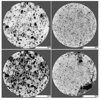
The samples before and after the reactive flow experiments were imaged in the dry state using a high resolution µ-CT scanner at the Australian National University (ANU)[24]with at least 8.5×8.5×8.5 mm field of view and resolutions of less than 5 µm.The post-dissolution image was superposed to the pre-dissolution image using a 3D registration technique developed by Latham et al. [25].Figure 1 (top images) illustrates examples of registered slices through the 3D µ-CT images of the pre-dissolution oolitic and wackestone-packstonesamples at the resolution of 4.49 and 4.33 μm, respectively.
3. Results
Figure 1 shows examples of registered slices of the oolitic and wackestone-packstone samples before and after the dissolution experiments. Visual observations of the images illustrate the occurrences of two different dissolution regimes within the samples. In the case of oolitic limestone, the reactive fluid locally increased the pore diameters across most parts of the sample (quasi-uniform dissolution), while the reactive fluid was consumed over small parts of the mineral surface area of the wackestone-packstone carbonate sample leading to the formation of a few highly conductive flow channels (wormholes dissolution).
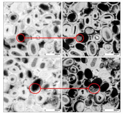
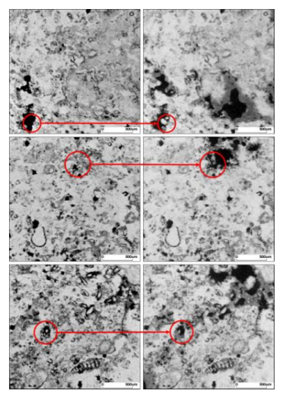
The precision of the registered images allowed us to observe local mineral detachment and displacement deposition in the pore space after the reactive flow experiments. Figures 2 and 3 illustrate the occurrence of regions (indicated by the red circles) showing voxels (or part of voxels) which are initially in the pore space but will finally be occupied by precipitated or mobilized mineral particles.
4. Discussion
Similar findings related to local particle displacement and deposition at the pore scale have been reported by other studies mainly using the µ-CT and SEM methods [6-8,26]. Garing, et al. [27] and Mangane, et al. [28] clearly showed areas within the samples where pores were filled by solid particles after the experiments. Beside the qualitative results, it is of great importance to quantitatively examine particle displacement/deposition as well as solid dissolution from the acquired images.
Qajar and Arns [5] have developed a method based on the 2D intensity histogram of registered images to compute local as well as overall dissolution and deposition fractions and also to distinguish between different dissolution patterns. It is worth mentioning that small volume regions suspected to mineral dissolution or deposition may be misinterpreted due to image artefacts, including image noise in particular. The uncertainty associated with this kind of problem was quantitatively studied by Qajar, et al. [23] by introducing a signal-difference-to-noise ratio (SDNR) parameter which is computed from pre- and post-dissolution image intensities. They argued that the SDNR values represent the relative importance of the actual density changes to the noise.
5. Conclusion
As found in this study, the µ-CT technique can provide high resolution images of successively disturbed samples. The images can be used to qualitatively investigate local changes within the samples including particle displacement and deposition. In addition, the registered images provide an important basis to calculate the evolution of the structure of porous media using advanced numerical techniques at the pore scale. The µ-CT is also a useful method to study pore scale reactive and non-reactive displacement mechanisms. For future directions, further investigations are needed (1) to evaluate the potentials of the µ-CT method to quantitatively analyse particle displacement within porous media, (2) to assess the uncertainty associated with the quantitative analysis of the image-based data, and (3) to relate the displacement of particles and possible pore clogging to macroscopic properties of the porous media.
References
- Arns, C.H., F. Bauget, A. Ghous, A. SakellarioU, T.J. Senden, A.P. Sheppard, R.M. Sok, W.V. Pinczewski, J.C. Kelly, and M.A. Knackstedt, Digital core laboratory: Petrophysical analysis from 3D imaging of reservoir core fragments. Petrophysics, 2005. 46(4): p. 260-277.
- Bernard, D., D. Gendron, J.-M. Heintz, S. Bordère, and J. Etourneau, First direct 3D visualisation of microstructural evolutions during sintering through X-ray computed microtomography. Acta Materialia, 2005. 53(1): p. 121-128.
- Gouze, P. and L. Luquot, X-ray microtomography characterization of porosity, permeability and reactive surface changes during dissolution. Journal of Contaminant Hydrology, 2011. 120-121: p. 45-55.
- Knackstedt, M.A., C.H. arns, A. Ghous, A. Sakellariou, T.J. Senden, A.P. Sheppard, R.M. Sok, H. Averdunk, W.V. Pinczewski, G.S. Padhy, and M.A. Ioannidis, 3D Imaging and Flow Characterization of the Pore Space of Carbonate Core Samples, in Paper SCA2006-23, presented at the International Symposium of the Society of Core Analysts. 2006, 12-16 September: Trondheim, Norway.
- Qajar, J. and C.H. Arns, Characterization of reactive flow-induced evolution of carbonate rocks using digital core analysis- part 1: Assessment of pore-scale mineral dissolution and deposition. Journal of Contaminant Hydrology, 2016. 192: p. 60-86.
- Lebedeva, E., T.J. Senden, M.A. Knackstedt, and N. Morrow, Improved Oil Recovery From Tensleep Sandstone- Studies of Brine-Rock Interactions by Micro-CT and AFM, in 15th European Symposium on Improved Oil Recovery. 2009, 27-29 April: Paris, France.
- Kumar, M., E. Lebedeva, Y. Melean, M. Madadi, A.P. Sheppard, T.K. Varslot, A.M. Kingston, S.J. Latham, R.M. Sok, A. Sakellariou, C.H. Arns, T.J. Senden, and M.A. Knackstedt, Micro-Petrophysical Experiments Via Tomography and Simulation, in Advances in Computed Tomography for Geomaterials. 2010, John Wiley & Sons, Inc. p. 238-253.
- Noiriel, C., D. Bernard, P. Gouze, and X. Thibault,Hydraulicpropertiesand microgeometry evolution accompanying limestone dissolution by acidic water. Oil & Gas Science and Technology-Rev. IFP, 2005. 60(1): p. 177-192.
- Cai, R., W.B. Lindquist, W. Um, and K.W. Jones, Tomographic analysis of reactive flow induced pore structure changes in column experiments. Advances in Water Resources, 2009. 32: p. 1396-1403.
- Ehrlich, R., S.K. Kennedy, S.J. Crabtree, and R.L. Cannon, Petrographic image analysis; I, Analysis of reservoir pore complexes. Journal of Sedimentary Research, 1984. 54(4): p. 1365-1378.
- Bekri, S., K. Xu, F. Yousefian, P.M. Adler, J.F. Thovert, J. Muller, K. Iden, A. Psyllos, A.K. Stubos, and M.A. Ioannidis, Pore geometry and transport properties in North Sea chalk. Journal of Petroleum Science and Engineering, 2000. 25(3-4): p. 107-134.
- Krekeler, M.P.S., E. Hammerly, J. Rakovan, and S. Guggenheim, Microscopy Studies of the Palygorskite-to-smectite Transformation. Clays and Clay Minerals, 2005. 53(1): p. 92-99.
- Hoefner, M.L. and H.S. Fogler, Pore Evolution and Channel Formation during Flow and Reaction in Porous-Media. AIChE Journal, 1988. 34(1): p. 45-54.
- Ketcham, R.A. and W.D. Carlson, Acquisition, optimization and interpretation of X-ray computed tomographic imagery: applications to the geosciences. Computers & Geosciences, 2001. 27: p. 381-400.
- Sakellariou, A., T.J. Sawkins, T.J. Senden, C.H. Arns, A. Limaye, A.P. Sheppard, R.M. Sok, M.A. Knackstedt, W.V. Pinczewski, L.I. Berge, and P.-E. Øren, Micro-CT Facility For Imaging Reservoir Rocks At Pore Scales, in paper presented at the SEG Annual Meeting, Society of Exploration Geophysicists. 2003: Dallas, Texas.
- De Beer, F.C. and M.J. Radebe, Neutron Imaging –Practice and Role as a Complementary NDE Technique to X-ray Imaging, in 18th World Conference on Nondestructive Testing, 16-20 April. 2012: Durban, South Africa.
- Kubis, A.J., G.J. Shiflet, D.N. Dunn, and R. Hull, Focused ion-beam tomography. Metallurgical and Materials Transactions A-Physical Metallurgy and Materials Science, 2004. 35A(7): p. 1935-1943.
- Fredrich, J.T., B. Menendez, and T.F. Wong, Imaging the Pore Structure of Geomaterials. Science, 1995. 268(5208): p. 276-279.
- Spanne, P., J.F. Thovert, C.J. Jacquin, W.B. Lindquist, K.W. Jones, and P.M. Adler, Synchrotron Computed Microtomography of Porous Media: Topology and Transports. Physical Review Letters, 1994. 73(14): p. 2001-2004.
- Sheppard, A.R., R.M. Sok, H. Averdunk, V.B. Robins, and A. Ghous, Analysis of Rock Microstructure Using High Resolution X-ray Tomography, in Paper SCA2006-26, presented at the International Symposium of the Society of Core Analysts. 2006, 12-16 September, 2006: Trondheim, Norway.
- Knackstedt, M.A., A. Golab, A. Carnerup, T. Senden, A. Butcher, and L. Riepe, Multi-scale Formation Evaluation of Tight Gas Resources, in Paper IPTC 14919-MS, presented at the International Petroleum Technology Conference, Society of Petroleum Engineers. 2011, International Petroleum Technology Conference: Bangkok, Thailand, doi:10.2523/ 14919-MS.
- Shah, S.M., F. Gray, J.P. Crawshaw, and E.S. Boek, Micro-computed tomography pore-scale study of flow in porous media: Effect of voxel resolution. Advances in Water Resources, 2015.
- Qajar, J., N. Francois, and C. Arns, Microtomographic Characterization of Dissolution-Induced Local Porosity Changes Including Fines Migration in Carbonate Rock. SPE J, 2013. 18(3): p. 545-562.
- Sakellariou, A., T.J. Senden, T.J. Sawkins, M.A. Knackstedt, M.L. Turner, A.C. Jones, M. Saadatfar, R.J. Roberts, A. Limaye, C.H. Arns, A.P. Sheppard, and R.M. Sok. An x-ray tomography facility for quantitative prediction of mechanical and transport properties in geological, biological and synthetic systems. in Developments in X-Ray Tomography IV. 2004.
- Latham, S., T. Varslot, and A.P. Sheppard, Image Registration: Enhancing and Calibrating X-ray Micro-CT Imaging, in Paper SCA2008-35, presented at the International Symposium of the Society of Core Analysts. 2008, 29 October-2 November, 2008: Abu Dhabi, UAE.
- Fogden, A., M. Kumar, N.R. Morrow, and J.S. Buckley, Mobilization of Fine Particles during Flooding of Sandstones and Possible Relations to Enhanced Oil Recovery. Energy & Fuels, 2011. 25(4): p. 1605-1616.
- Garing, C., P. Gouze, M. Kassab, M. Riva, and A. Guadagnini, Anti-correlated Porosity– Permeability Changes During the Dissolution of Carbonate Rocks: Experimental Evidences and Modeling. Transport in Porous Media, 2015. 107(2): p. 595-621.
- Mangane, P.O., P. Gouze, and L. Luquot, Permeability impairment of a limestone reservoir triggered by heterogeneous dissolution and particles migration during CO2-rich injection. Geophysical Research Letters, 2013. 40(17): p. 4614-4619.