


|
|
DOI Prefix 10.20431 |
Information
Journal Policies
A Rare Case of Bilateral Renal Lymphoma on FDG PET/CT Imaging
Pelin Ozcan Kara1,Zehra Pinar Koc2,Eylem Sercan Ozgur3,Erhan Ayan4,Anil Tombak5
2.Mersin University, Faculty of Medicine, Department of Chest Disease, Mersin, Turkey.
3.Mersin University, Faculty of Medicine, Department of Thoracic Surgery, Mersin, Turkey.
4.Mersin University, Faculty of Medicine, Department of Haematology, Mersin, Turkey.
Copyright : © 2017 . This is an open access article distributed under the Creative Commons Attribution License, which permits unrestricted use, distribution, and reproduction in any medium, provided the original work is properly cited.
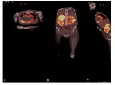
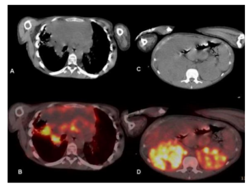
Twenty-seven years old women with suspected malignancy of anterior mediastinal mass underwent PET-CT imaging for metabolic charecterisation and initial staging. F-18 FDG PET/CT demonstrated anterior mediastinal hypermetabolic necrotic mass with cervical, mediastinal, intraabdominal hypermetabolic lymphadenopaties. Although, FDG is excreted through kidneys diffuse and multifocal renal FDG uptake in enlarged kidneys were observed. We report a patient presenting with acute renal failure and suspected malignancy. The diagnosis of diffuse renal involvement in lymphoma is important on FDG PET/CT imaging and always abnormal.
Lymphoma, renal lymphoma, FDG PET/CT,Radiology and Medical Imaging
1. Introduction
Extra nodal involvement areas such as gastrointestinal, head and neck, orbital, central and peripheral nervous system, thorax, bone, skin, breast, testis, thyroid and genitourinary system can be seen in 25-40% of HL and especially NHL patients, although it is known as lymph node malignancy [1,2]. Extra nodal involvement is important in terms of prognosis. Imaging procedures play a very important role in diagnosis. 18F-FDG PET-CT imaging is a standard method in lymphoma patients and has a special importance in these patients in terms of superiority of CT imaging in detecting extra nodal regions. The role of 18F-FDG PET-CT in detection, staging and restaging of patients with extranodal involvement in NHL has also been reported in the literature [3,4]. In this case presentation, in addition to nodal and mediastinal involvement areas in a patient diagnosed as NonHodgkin lymphoma, rare bilateral renal extra nodal simultaneous involvement detected on 18F-FDG PET-CT imaging were presented.
2. Case Report
A 27-year-old woman with mediastinal mass suspected for malignancy with nondiagnostic two biopsy results observed on diagnostic CT imaging was directed to our department for performing PET-CT imaging for metabolic characterisation, for finding appropriate biopsy region and initial staging. Following 6 hours of fasting, while the patient had a blood glucose level of 90 ml/ dL, whole-body PET-CT imaging with low-dose nondiagnostic CT in 3D mode was performed 60 min later after injection of 10 mCi (370 MBq) i.v. 18-F-FDG. On PET/ CT imaging (GE Discovery PET-CT 610),hyper metabolic (SUVmax: 2-9) lymphadenopathies were observed in left servical level IV and supraclavicular, mediastinal, mesenteric, retroperitoneal and renal hilus region. An anterior mediastinal hyper metabolic (SUVmax: 14.05) necrotic mass starting from thyroid lob and extending inferiorly, surrounding mediastinal vascular structures was also observed. In addition; So many hyper metabolic (SUVmax: 12.9) nodular lesions in bilateral enlarged kidneys with diffuse FDG uptake was detected. According to PET/CT findings the diagnosis of lymphoma was first considered. Before FDG PET/CT imaging, because of nondiagnostic biopsy results from necrotic mediastinal mass, renal biopsy was recommended. She diagnosed as nonHongkin Lymphoma (DLBCL) after renal biopsy.
3. Discussion
Extranodal lymphoma originating from solid organs accounts for approximately one third of NHL patients. Kidney involvement can also be seen often [5]. By contrast, primary renal lymphoma without other organ or nodal involvement is a rare disease [6-8]. In the current case, the patient had acute renal failure as initial presentation and kidneys were extranodal lymphoma regions with mediastinal and nodal involvement. Despite the high frequency of renal involvement in DLBCL as an extranodal site, simultaneous involvement of both kidneys is very rare. On FDG PET/CT imaging enlargement of both kidneys without obstruction, diffuse and multifocal FDG uptake were observed. Differention of primary and secondary renal lymphoma may be complicated by imaging procedures. Diagnosis can be established unexpectedly following radical nephrectomy, since the tumors are initially assumed to be renal cell carcinoma [9]. Diffuse FDG renal uptake is reported in literature in few reports [10,11]. A recent study evaluated the ability of PET/CT to differentiate renal cell carcinoma (RCC) from renal lymphomatous involvement from Nicolau C. et al. [12]. In this case presentation, in addition to nodal and mediastinal involvement areas in a patient diagnosed as NonHodgkin lymphoma, rare bilateral renal extra nodal simultaneous involvement detected on 18F-FDG PET-CT imaging were presented. PET/CT was found useful for demonstrating biopsy site and also differentiation of primary renal lymphoma versus extra nodal involvement.
4. Conclusion
The diagnosis of diffuse renal involvement in lymphoma is important on FDG PET/CT imaging and always abnormal.
References
- Lopez-Guillermo A, Colomo L, Jimenez M, Bosch F, Villamor N, Arenillas L, et al. Diffuse large B-cell lymphoma: clinical and biological characterization and outcome according to the nodal or extranodal primary origin. J Clin Oncol. 2005;23:2797–804.
- Economopoulos T, Papageorgiou S, Rontogianni D, Kaloutsi V, Fountzilas G, Tsatalas C, et al. Multifocal Extranodal Non- Hodgkin Lymphoma: α clinicopathologic study of 37 Cases in Greece, a Hellenic Cooperative Oncology Group Study. Oncologist. 2005;10:734–8.
- Paes FM, Kalkanis DG, Sideras PA, Serafini AN. FDG PET/CT of Extranodal Involvement in Non-Hodgkin Lymphoma and Hodgkin Disease. RadioGraphics. 2010;30:269–91.
- Even-Sapir E, Lievshitz G, Perry C, Herishanu Y, Lerman H, Metser U. Fluorine-18 Fluorodeoxyglucose PET/CT Patterns of Extranodal Involvement in Patients with Non-Hodgkin Lymphoma and Hodgkin’s Disease. Radiol Clin N Am. 2007;45:697–709.
- Cabuk D, Gullu YT, Basyigit I, Acikgoz O, Uygun K, Yildiz K and Yildiz F: Multifocal extranodal involvement of diffuse large B-Cell lymphoma. Case Rep Pulmonol 2013: 794642, 2013.
- Pahwa M, Gupta N, Tyagi V and Chadha S: Primary renal lymphoma: Is prognosis really that bad? Saudi J Kidney Dis Transpl 24: 816-817, 2013.
- Hart S, Ellimoottil C, Shafer D, Mehta V and Turk TM: A case of primary renal lymphoma. Urology 80: 763-765, 2012.
- Torrecilla García-Ripoll JR, Pascual Samaniego M, Martín Blanco S, Rivera Ferro J, Peral Martínez JI and Fernández del Busto E: Primary renal lymphoma. Actas Urol Esp 27: 555-558, 2003 (In Spanish).
- Valarmathi K, Jamila A, Ravi S, Selvambigai and Muthulatha: A rare case of renal tumour. J Clin Diagn Res 7: 2006-2007, 2013.
- Navalkissoor S, Szyszko T, Gnanasegaran G, Nunan T. Diffuse FDG renal uptake in lymphoma. Clin Nucl Med. 2010 Oct;35(10): 813-5.
- Tan YZ, Yılmaz S, Özhan M, Halaç M. FDG PET-CT Finding in Bilateral Renal and Bone Involvement of Diffuse Large B-Cell Lymphoma. Mol Imaging Radionucl Ther. 2014 Oct 5;23(3):104-6.
- Nicolau C, Sala E, Kumar A, Goldman DA, Schoder H, Hricak H, Vargas HA. Renal Masses Detected on FDG PET/CT in Patients With Lymphoma: Imaging Features Differentiating Primary Renal Cell Carcinomas From RenalLymphomatous Involvement. AJR Am J Roentgenol. 2017 Apr;208(4):849-853.