


|
|
DOI Prefix 10.20431 |
Information
Journal Policies
The Tip of the Ice-Berg: A Case of an Impacted Third Molar Fusion
Geon Pauly1,Roopashri Rajesh Kashyap2,Raghavendra Kini3,Prasanna Kumar Rao4,Gowri P Bhandarkar5,Ujwala Shetty6
2.Reader, Department of Oral Medicine and Radiology, A J Institute of Dental Sciences, Kuntikana, Mangaluru, Karnataka, India.
3.Professor and Head, Department of Oral Medicine and Radiology, A J Institute of Dental Sciences, Kuntikana, Mangaluru, Karnataka, India.
4.Professor, Department of Oral Medicine and Radiology, A J Institute of Dental Sciences, Kuntikana, Mangaluru, Karnataka, India.
5.Assistant Professor, Department of Oral Medicine and Radiology, A J Institute of Dental Sciences, Kuntikana, Mangaluru, Karnataka, India.
Copyright : © 2017 . This is an open access article distributed under the Creative Commons Attribution License, which permits unrestricted use, distribution, and reproduction in any medium, provided the original work is properly cited.
Odontogenic anomalies of teeth are found at large in our day to day practice. Developmental anomalies of number such as fusion and gemination are often encountered clinically and can only be distinguished after radiographic interventions in most cases. Tooth fusion are seen in erupted teeth predominantly, thus making fusion of impacted teeth comparatively rare. This article highlights the importance of clinical and radiographic correlation in arriving at a definitive diagnosis.
Ice-Berg,Impacted Third Molar Fusion,Radiology and Medical Imaging
1. Case Report
A 21-year-old male had reported to our department with throbbing pain in the lower left back tooth region since 10-15 days. His medical and dental histories were non-contributory. On examination of the left side, there was presence of a tilted cusp-tip of the mandibular third molar visible distal to second molar. A radiograph was advised suspecting an impacted third molar.
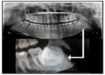
IOPA was not possible ailing to patient’s discomfort during the procedure, so a panoramic radiograph was taken. The panoramic image revealed a mesio-angularly tilted rather alarmingly bulbous crown with accessory number of cusps, while the roots were distinctly separate suggesting that the altered morphology was because of a fusion between single rooted third molar and possibly a disto-molar [Figure 1]. The patient was referred to department of oral surgery for surgical removal of the tooth. Dis-impaction was carried out under local anaesthesia. There were no associated post-surgical complications.
2. Discussion
Fusion of primary or permanent teeth are rare developmental anomalies that stems from the embryogenic union of two teeth originating from two or more tooth germs and can be seen between two adjacent teeth or a tooth and supernumerary teeth as in our case [1]. The aetiology is still unclear at large. However, physical force or pressure producing close contact between two developing tooth buds has been reported as a possible cause. Also, genetic, various idiopathic environmental factors and even trauma have been considered as contributing factors [2]. The terms Twinning, Joined tooth or Double tooth, is used to describe both fusion and germination making accurate diagnosis a challenging task. Two tooth’ rule was introduced in 1979 to differentiate fusion and germination. If the fused tooth is considered as one and the number of teeth in the dental arch is less, then the term fusion is considered. If the number of teeth in the dental arch are normal, then it is termed as gemination or it is a case of fusion between normal and supernumerary as seen in our case [3].
Depending on factors such as location of the connecting area, root-development stage, patient age and clinical symptoms, treatment of fused teeth may vary. Treatment for symptomatic cases range from extraction of concerned tooth, root-canal therapy, to removal of the only the unwanted part, and its eventual re-implantation into its original site is also an approach less common but successfully attempted [4].
3. Conclusion
Developmental anomalies such as tooth fusion have been found as an aesthetic barrier or even as a dormant trouble-maker just waiting to be discovered as in our case. It is therefore extremely essential to undertake the right radiographic investigations before surgical procedures to accurately diagnose and thus effectively carry out the required procedure.
References
- A. Knežević, S. Travan, Z. Tarle, J. Šutalo, B. Janković, and I. Ciglar, “Double tooth,” Collegium Antropologicum, 2002; 26(2): 667– 72.
- Crawford NL, North S, Davidson LE. Double permanent incisor teeth: management of three cases. Dental update. 2006; 33(10): 608–610.
- Rao PK, Mascarenhas R, Anita A, Devadiga D. Fusion in Deciduous Mandibular Anterior Teeth – A Rare Case. Dentistry. 2014; 2: 001.
- Guler DD, Tunc ES, Arici N, Ozkan N. Multidisciplinary Management of a Fused Tooth: A Case Report. 2013; 1(2): 1-5.