


|
|
DOI Prefix 10.20431 |
Information
Journal Policies
ARC Journal of Ophthalmology
Volume-3 Issue-2, 2018, Page No: 11-12
Epiphora Decoded-A Rare Case Report
Dr. Anubhav Chauhan M.S
Senior Resident, Department of Ophthalmology, Dr. Yashwant Singh Parmar Govt. Medical College, Nahan, District Sirmour, Himachal Pradesh, India.
Citation : Dr. Anubhav Chauhan M.S, "Epiphora Decoded-A Rare Case Report" ARC Journal of Ophthalmology. 2018; 3(2): 11-12.
Copyright : © 2018 . This is an open access article distributed under the Creative Commons Attribution License, which permits unrestricted use, distribution, and reproduction in any medium, provided the original work is properly cited.
Abstract
Epiphora, Plastic, Punctum,Ophthalmology
We report a case of a 28-year-old female with a history of persistent watering in the right eye. A retained plastic strip in the lacrimal punctum proved to be the culprit. Failure to detect the causative agent can lead to unnecessary use of drugs not needed. It is a rare case report.
Epiphora, Plastic, Punctum,Ophthalmology
1. Introduction
A foreign body in the lacrimal punctum can often be missed leading to unnecessary delay in proper treatment leading to chronic ocular irritation and watering. Sometimes, these cases are wrongly diagnosed as having conjunctivitis and start receiving treatment for the same, while the culprit i.e foreign body is still lodged in the punctum. Herein, we describe a very rare case of plastic strip in the lacrimal punctum.
2. Case
A 28-year-old female (figure 1) presented with a history of watering and redness in the right eye since last three days. History revealed that she had developed the symptoms while collecting wood in the area around her home. Bilateral visual acquity was 6/6, with a normal fundus on examination.
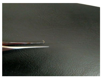
Slit-lamp examination revealed a tiny plastic strip (figure 2) lodged in the lower lacrimal punctum in the right eye. This was removed with a forceps. No other ocular trauma was noted. There was no previous history of ocular illness or surgery. Patient was given topical antibiotics and lubricants. All the symptoms subsided subsequently by the next day.
Figure1.
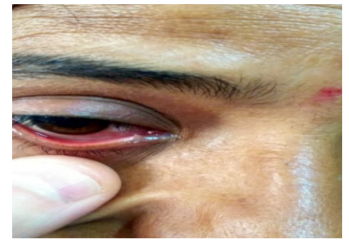
Figure2.
3. Discussion
Retained intraocular foreign bodies can present as unilateral conjunctivitis/ kerato conjunctivitis[1]. Cilia have been reported to be found embedded in the lacrimal puncta,[2] meibomian gland orifice, subconjunctival space and even corneal stroma. The patient usually presents with a history of foreign body sensation and watering[3].
Eyelashes are reported to enter the upper punctum much more frequently as compared to the lower punctum[4]. Our case highlight the importance of careful clinical examination of the lids to prevent misdiagnosis and a subsequent wrong treatment. Protective glasses are advised for people working in environment where ocular injuries are prone. To the best of our knowledge, this is the first report wherein a plastic material has been reported to be embedded in the lacrimal punctum.
References
- Kalavathy CM, Parmar P, Kaliamurthy J, Jesudasan CA, Thomas PA. Keratoconjunctivitis caused by an unusual retained conjunctival foreign body: a frequently unrecognized entity. Indian J Ophthalmol 2014; 62(5): 633–5.
- Singh S, Narang P, Mittal V. Getting hooked: Eyelash in lacrimal punctum. Saudi Journal of Ophthalmology 2017; 31(3): 201–2.
- Meel R, Vashisht S. Eyelash in Lacrimal Punctum.Delhi Journal of Ophthalmology 2013; 23(3): 227.
- Nagashima K, Kido R. Relative roles of upper and lower lacrimal canaliculi in normal tear drainage. Jpn J Ophthalmol 1984; 28:259-62.