


|
|
DOI Prefix 10.20431 |
Information
Journal Policies
Conservative Management of Congenital Dacryocystocele Complicated With Dacryocystitis and Cellulitis
Corredor-Osorio Rafael*, Buitrago-Corredor Vanessa Gabriela
Copyright : © 2018 . This is an open access article distributed under the Creative Commons Attribution License, which permits unrestricted use, distribution, and reproduction in any medium, provided the original work is properly cited.
Congenital dacryocystoceles are a variant of nasolacrimal duct obstruction. They usually have a bluish color and are located around the medial canthal area. Early treatment of dacryocystoceles is indicated. The treatment is controversial, some authors recommend a conservative medical management, and others a surgical intervention. Congenital dacryocystoceles with secondary dacryocystitis in the newborns may develop. Here, we report a case of congenital dacryocystocele which was managed with topical antibiotic local and lacrimal massage. Four days after his first visit to our medical center, the patient returned presenting an infection (acute dacryocystitis and cellulitis) which was treated with oral antibiotics and warm compresses.
Complete resolution was observed in the patient and no recurrence was noted upon after examination ten days later. The diagnosis of congenital dacryocystocele can be made on clinical findings alone.
We suggest that patients with dacryocystocele without secondary infection shoul d be treated with topical antibiotics and massage. If possible infection is detected, an oral antibiotic and warm compresses should be added with a careful observation.
Dacryocystocele; dacryocystitis; cellulitis; nasolacrimal duct obstruction.
1. Introduction
The congenital dacryocystocele is an uncommon malformation consisting of an obstruction of the lacrimal excretory system, mainly in the Hasner valve (in the nasolacrimal ostium) or Rosenmüller's (between the common duct and the lacrimal sac), an accumulation of mucous secretion to that level with its consequent dilation [1], with an incidence of 0.02 % in newborns [2], it is usually unilateral [2-5], it has a familial predisposition, and it is more frequent in females [2-5].
Dacryocystocele, which usually noticed in the first weeks after birth [1,3] as a blue-colored cystic mass over the lacrimal sac [2,4]. The lesions may resolve spontaneously,[4] but secondary dacryocystitis may occur,[5,6] however the treatment for congenital dacryocystoceles is controversial [3,4,6]. Some authors recommended conservative treatment [4,5,7] (lacrimal massage, warm compresses, topical and/or systemic antibiotic) and others authors surgical intervention, such as nasolacrimal duct probing [1,2] or nasal cyst marsupialization [2,5,6].
As possible complications, secondary dacryocystitis can develop. If untreated, it could complicate by a progressive cellulitis and formation of an abscess within the lacrimal sac [3] and when these dacryocystoceles extend intra- nasally and form a nasal cyst in the inferior meatus causing blockage of the nasal cavity can cause respiratory compromise [3, 6].
The differential diagnosis includes other masses such as dermoid and epidermoid cyst, hemangioma, glioma, encephalocele, tumor, lymphangioma. [1,3,7] In this case report, we presented an 8 days-old newborn with unilateral congenital dacryocystocele complicated with dacryocystitis and cellulitis preseptal. This condition probably resolves spontaneously but, we suggest a conservative treatment with careful observation of any signs of infection.
2. Case Report
An 8-days-old infant presented with the complaint a bluish cyst mass, just inferior the right medial canthal area. He had been born healthy.
External examination revealed an approximately 1.0 cm circumscribed soft, cyst non tender swelling was present in the right lacrimal sac region. The swelling was presented just inferior and lateral to the right medial canthus (Figure 1). The overlying skin was minimally erythematous and the mass of cyst firm consistency. The mass was free from de overlying skin. Both puncta were present. Compression of the lacrimal sac area did not show any regurgitation of fluid. Rest of the anterior segment and retinal examinations were within normal limits.
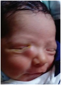
Based on these clinical findings, a diagnosis of unilateral congenital dacryocystocele was made. The patient was treated with topical tobramycin sulphate (Tobrasol solution, LO Oftalmi Venezuela) and lacrimal massage. Computed tomography scan (CT) is proposed to confirm the diagnosis. An examination under anesthesia and probing of the lacrimal system was planned for one week later.
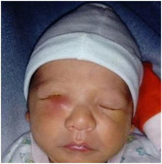
4 days later, the mother reported progressive erythema and swelling around the cyst (Figure 2). We continued the conservative treatment, but added a daily administration of oral Amoxicillin+ Clavulanate (Augmentin 200 mg, Glaxo Smith Kline drug) half dosage twice a day for 10 days with warm compresses and careful observation. Two days later, the mother reported that the prior night, there had been yellowish discharge from the right eye, and the swollen, blue cyst area had spontaneously decompressed. Massage, warm compresses, eye drops and oral antibiotics administration were continued. Given, that the dacryocystocele had spontaneously, decompressed, the CT scan and probing of nasolacrimal system was deferred. The condition the resolved after four days (Figure 3).

3. Discussion
Dacryocystocele, also known as amniocele, amniotocele, or dacryocele, are a uncommon form of congenital nasolacrimal duct obstruction with both proximal and distal obstruction resulting in lacrimal sac enlargement. Usually presenting at birth, there is significant risk for infection [8]. The management of dacryocystocele is controversial [2-4, 6]. Many of cases of dacryocystocele resolve spontaneously, [3,4,9] or with conservative treatment, and it includes antibiotic drops, lacrimal massage and warm compresses [8].
We followed the conservative treatment protocol, but our patient presented days later a dacryocystitis with cellulitis and we prescribed oral antibiotics and careful observation. Two days later, a spontaneous resolution of the dacryocystocele was presented. Mimura et al [4] explain that this spontaneous resolution is perhaps due to the proximal obstruction was opened before the distal obstruction, the dacryocystocele will rupture to the common canaliculus, draining the mucoid liquid through the puncta. This also explains that there is a lacrimal passage from the lacrimal sac to the lacrimal system that could lead to a dacryocystitis. With our conservative treatment, we found that the patient developed dacryocystitis and cellulitis but when adding an oral antibiotic and compresses, there was a rupture of the dacryocystocele with drainage of mucoid fluid. Symptoms resolved with topical and systemic antibiotics without surgical intervention. This finding suggests that there is a possibility that the rupture of the dacryocystocele may occur, while the neonate presented with dacryocystitis. Schnall and Christian reported that 76% of the dacryocystoceles in their study resolved within 2 weeks with conservative treatment protocol consisting of warm compresses and massage. There was no need for intravenous antibiotics and computed tomography.
We suggest conservative treatment for dacryocystoceles that have a high probability of spontaneous resolution with a topical antibiotic, warm compresses, massage and careful observation of any infectious signs. Surgical intervention such as nasolacrimal duct probing will be performed only if medical management failed [1-3,8] .
Infected dacryocystoceles and cellulitis should be treated with an efficient use of topical and systemic antibiotics, warm compresses, and massage [3, 8, 10].
4. Consent
Written informed consent was obtained from the patient’s parents for publication of this Case Report and any accompanying images.
5.References
- Machado MA., Abreu Junior Ld., Silva JA., Allemann N. Congenital dacryocystocele : diagnosis using ante and post-natal ultrasonography. Arq Bras Oftalmol 2014; 77(4): 261-263.
- Shekunov J, Griepentrog GJ., Diehl NN., Mohney BG. Prevalence and clinical characteristics of congenital dacryocystocele. JAAPOS 2010; 14(5): 417-420. DOI:10.1016/ jaapos.2010.0.006.
- Lueder GT. The association of neonatal dacryocystoceles and infantile dacryocystitis with nasolacrimal ducts cysts (an American ophthalmological society thesis), Trans Am Ophthalmol Soc 2012; 110:74-93.
- Mimura M., Ueki M., Oku H., Sato B. et al. Process of spontaneous resolution in the conservative management of congenital dacryocystocele.
- Cavazza S. Laffi GL., Lodi L., Tassinari G. et al. Congenital dacryocystocele: diagnosis and treatment. Acta Otorhinolaryngol Ital 2008; 28: 298-301.
- Ismi O., Bozkurt FM., Icme G. Eti C. et al. A rare cause of intermittent respiratory distress and epiphora in the newborn: congenital dacryocystocele. Gland Surg 2017; 6(1): 114- 118.
- Ha YJ. Choi HY. Myung KB. Choi YW. A case of congenital dacryocystocele. Ann Dermatol 2010; 22(1): 54-56. Doi: 10.5021/ ad.2010. 22.1.54.
- Wasserman BN, Schnall BM, Levin AV. Sequential bilateral dacryocele. Arch Ophthalmol 2011; 12(1): 104-105.
- Geenlaw SM, Chaney KS, Belazarian L, Wiss K. Congenital dacryocystocele. J Am Acad Dermatol 2009; 61(6): 1088-1090.
- Schnall BM, Christian CJ. Conservative treatment of congenital dacryocele. J Pediatr Ophthalmol Strabismus . 1996; 33(5): 219–222.