


|
|
DOI Prefix 10.20431 |
Information
Journal Policies
Percutaneous Transluminal Embolisation of Intraparenchymal
Pseudoaneurism and High Flow Arteriovenous Fistula Developed After Biopsy in Renal Allograft: Case Report
Sultan Ozkurt1*, Berat Acu2, Elif Gundogdu2, Cigdem Mengus1, Ahmet Ugur Yalcin1
2.Department of Radiology, Faculty of Medicine, Eskisehir Osmangazi University, Eskisehir, Turkey.
Copyright : © 2018 . This is an open access article distributed under the Creative Commons Attribution License, which permits unrestricted use, distribution, and reproduction in any medium, provided the original work is properly cited.
In transplantation practice percutaneous needle biopsy is very important in renal allograft management. After biopsy, vascular complicatons like AVF and pseudoaneurism can occur rarely and because of they are generally asymptomatic their diagnosis could delay. We detected an intraparenchymal giant pseudoaneurism and high flow AVF in renal allograft while we search for etiology of hypertension and deterioration of renal functions for 2 months in 27 years old woman patient who has history of renal biopsy 7 years ago and renal transplantation from live donor 9 years ago. Due to presence of rupture risk, renal hypertension and dysfunction, vascular lesion was treated successfully with percutaneous transluminal embolisation. If transcutaneous needle biopsy was applied to renal allograft and there is a clinical symptom or vascular complication finding, imaging of allograft with Color Duplex Doppler ultrasonography (US) should be done periodically.
arteriovenous fistula; percutaneous transluminal embolisation; pseudoaneurism,Nephrology
1. Introduction
Renal artery aneurisms are rarely seen. Their incidence is between %0,1-1 and %80 of them is in saccular form[1]. In transplantation practice, about this situation there are limited numbers of reports and they are about donor renal artery aneurism, mycotic aneurisms at vascular anastomosis line, that occur after transplantation or repairment of nonrenal vascular aneurisms[2-5].
Both in native or transplantated kidneys, vascular complication rate that occur after percutaneous core biopsy is between %0,9 - %15[6,7]. This wide difference is attributed to lesions are benign and asymptomatic[6]. There are not enough information about treatment indications and repairment technics of intraparenchymal aneurisms in renal allogrefts.
2. Case Description
A 27-year-old female patient was diagnosed with crescentic glomerulonephritis secondary end-stage renal disease and renal transplantation was performed 9 years ago from a live donor (mother).2 years after transplantation, renal biopsy was planned due to progression of 1.8 g/day proteinuria in patient who was treated with tacrolimus, mycofenolat mofetil and steroid based immunsupressive therapy and had totally normal renal functions for 2 years after transplantation. Despite of 5 attempts, no enough material was obtained and proteinuria regressed with irbesartan treatment (150 mg), renal functions were normal. At that time, serological examinations including BK virus were done to explain proteinuria, but no pathological finding was found.
At last 2 months, serum creatinin (SCr) levels were slightly increased (23.05.2018 SCr: 0.97 mg/dl, 20.07.2018 SCr: 1.28mg/dl) and the patient was hypertensive. At July 2018, in graft Color Duplex Doppler ultrasonography, 2.5 cm cyctic formation was detected at upper pole of transplantated kidney and there was ying- yang flow pattern in this formation. Next to it, arterialized flow patterned intrarenal vein and normal flow patterned intrarenal segmentary renal artery, whose flow speed was increased (peak systolic speed approximately 200 cm/sc) and resistance was decreased (RI:0.35), was detected.
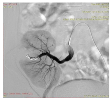
This findings were accepted significant for arterio venous fistula (AVF) with pseudoaneurism in the patient who had allograft biopsy before. The patient was hospitalized to confirm the diagnosis and for treatment. In her physical examination, blood pressure was 160/100 mm/Hg, SCr: 1.12 mg/dl, BKV DNA (urine) negative, takrolimus level: 5.4 ng/ml, in urine examination protein (+1), eryhtrocyte 1/HPF, leukocyte 1/HPF, spot urine protein/Cr ratio was 860 mg/day. She had taken tacrolimus (4 mg/day), mycofenolat (2gr/day), low dose prednisolone (5mg/day) and 10 mg amlodipin (irbesartan was changed into amlodipin for her pregnancy plans 2 years ago.) After giving fluids with N acetyl cystein to protect the kidney, selective renal angiography was done at 25.07.2018. A diagnostic angiography, followed by endovascular embolization of the fistula was planned. The procedure was carried out in the angiography suite of the interventional radiology unit. Following preparation of the left inguinal area under sterile conditions, a 6 French introducer sheat was placed into the left common femoral artery. A 5 French Cobra II catheter and a 0.035 inch hydrophilic guidewire were then inserted through the introducer sheath and the right iliac artery was catheterized using a contralateral approach. Angiographic images demonstrated the transplant kidney in the right iliac fossa, with its arterial and venous anastomoses to the right internal iliac artery and vein, respectively. The segmental branch of the renal artery supplying the middle part of the transplant kidney was increased in diameter. A high- flow AV fistula, with a giant aneurysmatic dilatation on its venous side, was visualized (figure 1). To reach the fistula site, the renal artery of the transplant kidney was then catheterized with a 5 French Cobra II catheter, and a 2.7 French co-axial microcatheter (Progreat Terumo) was selectively introduced through this catheter. Multiple pushable hydrocoils were delivered through the microcatheter, and the fistula site along with its arterial side, was embolized. Final angiographic images showed the complete occlusion of the fistula, without any aneurysmatic contrast filling (figure 2). No complications were observed during or after the procedure. The patient was discharged after 3 days of observation. At 03.08.2018 dated policlinic visit, we detected that blood pressure 110/70 mm/Hg without any antihypertensive drugs, SCr: 1.3 gr/dl, spot urine protein/Cr ratio: 298 mg/day.
Figure1: A diagnostic angiography
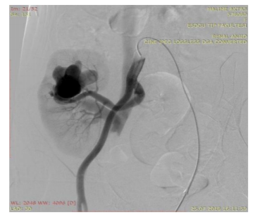
Figure2: After endovascular embolization
3. Discussion
AVF and pseudoaneurisms are iatrogenic damages which are related to biopsy in renal allografts and native kidneys. In our patient, because of having history of allograft biopsy and presence of intraparenchymal aneurism, aneurism is thought to be secondary to biopsy. In symptomatic patients; stent, coil and endovascular treatment via embolisation are used successfully in the treatment of these complications[8].
AVFs are attributed to simultaneous destruction of adjacent arterial and venous vessel walls and subsequent repair procedures. AVF frequency in allograft after renal biopsy is between %0.5 and %16 (9). %70 of AVF’s are asymptomatic and resolve spontaneously throughout weeks or months[10,11]. Pseudoaneurisms are caused by the bleeding that is triggered by destruction of arterial wall with biopsy. Then, during the repair processes of perivascular tissues, they liquefy and dissolve leaving a continuous sac[9]. Biopsy triggered AVF and pseudoaneurisms can be detected by Color Duplex Doppler ultrasonography which is able to detect intrarenal blood flow changes, their localization and dimension are defined with selective angiography[10,12]. Some writers suggest conservative therapy for biopsy related allograft damages[13,14]. Interventional radiology is supposed to be necessary when AVF and pseudoaneurisms cause severe clinical findings (like hematuria, high flow shunt, deterioration of renal functions), in case of high risk of rupture, severe hypertension, progressive volum increase of pseudoaneurism and diameter of more than 2.5 cm.10 Large pseudoanerisms have rupture risk without any intervention and in case of severe hypertension related to aneurism there is intervention indication[15]. When pseudoaneurisms enlarge over time, it can cause intrarenal blood flow restriction by pressure, therefore renal ischemia can lead to increase of blood pressure and deterioration of renal function as seen in our patient[15]. Finally, in our case aneurism was large and it could cause this type of pressure and regulation of blood pressure without any drug after intervention suggests the relation of high blood pressure with aneurism. Pseudoaneurism and AVF can be symptomatic by enlarging over time while they are small and asymptomatic at the beginning[16]. Nakatani et al. reported that they did a successful endovascular intervention to pseudoaneurism and AVF, diagnosed 5 years later from allograft biopsy .16 In the previous reports, the time between biopsy and intervention was mentioned between 2,9 days and 5 years[16,17]. Our case is different from other cases because of diagnosis of graft dysfunction, hypertension and vascular complication 7 years later from biopsy.
In conclusion, in renal transplant patients there is risk of iatrogenic pseudoaneurism and AVF despite the importance of percutaneous needle biopsy. Renal pseudoaneurisms can be detected after years and their aneurismal pressure effect can lead to allograft dysfunction and hypertension. It is precaution to evaluate allograft periodically with Color Duplex Doppler ultrasonography.
References
- Olakkengil S, RM Mohan. Transplantation of kidneys with renal artery aneurysm. Clinical Transplantation 2011; 25: 516–9.
- 2.Paloyo S, Sageshima J, Chen L, Burk GW, Ciancio G. Aneurysmectomy with Partial Nephrectomy on a Living Donor Renal Allograft: A Case Report. Case Reports in Transplantation 2013; 2013; 791413.
- Kountidou CS, Stier K, Niehues SM, Lingnau A, Schostak M, Fuller TF et al. Successful Repair of Post-transplant Mycotic Aneurysm of Iliac Artery With Renal Graft Preservation: A Case Report. Urology 2012; 80:1151-53.
- Laouad I, Buchler M, Noel C, Sadek T, Maazouz H, Westeel PF et al. Renal Artery Aneurysm Secondary to Candida albicans in Four Kidney Allograft Recipients. Transplantation Proceedings 2005: 37; 2834– 36.
- Kim HK, Ryuk JP, Choi HH, Kwon SH, Huh S. Abdominal aortic aneurysm repair in patient with a renal allograft: a case report. J Korean Med Sci 2009; 24:166-69.
- Gerald DDM, Ajit S, Albert BZ. Imaging of vascular complications associated with renal transplants. Am J Roentgenol 1991; 157:449–59.
- Selim M, Goldstein MJ. Case report: Parenchymal pseudoaneurysm of a renal allograft after core needle biopsy: A rare cause of allograft injury. Transplant Proc 2011; 43:2781–83.
- P.D. Orons, A.B. Zajko. Angiography and interventional aspects of renal transplantation.
- Radiol Clin North Am 1995; 33:61.
- Martinez T, Palomares M, Bravo JA, Alvarez G, Galindo P, Entrena AG et al. Biopsy-induced arteriovenous fistula and venous aneurysm in arenal transplant. Nephrol Dial Transplant 1998: 13:2937.
- Matsell DG, Jones DP, Boulden TF, Burton EM, Baum SL, Tonkin IL. Arteriovenous fistula after biopsy of renal transplant kidney: diagnosis and treatment. Pediatr Nephrol 1992: 6: 562.
- Beaujeux R, Boudjema K, Ellero B, Rimmelin A, Roy C, Dietemann JL et al. Endovascular treatment of renal allograft post-biopsy arteriovenousfistula with platinum microcoils. Transplantation 1994: 57:311.
- Bach AM, Merton DA, Burke JF Jr, Halpern EJ. Sonographic diagnosis of arteriovenous fistula and pseudoaneurysm after biopsy of a transplanted kidney. J Ultrasound Med 1993:12: 545.
- Hubsch P, Schurawitzki H, Traindl O, Karnel F. Renal allograft arteriovenous fistula due to needle biopsy with late onset of symptoms – diagnosis and treatment. Nephron 1991: 59: 482.
- Taylor KJ, Morse SS, Rigsby CM, Bia M, Schi M. Vascular complications in renal allografts: detection with duplex Doppler US. Radiology 1987:162:31.
- Glebova NO, Brooke BS, Desai NM, Lum YW. Endovascular interventions for managing vascular complication of renal transplantation. Semin Vasc Surg 2013; 26:205–12.
- Nakatani T, Uchida J, Han YS, Iwai T, Nakamura K, Kawashima H et al. Renal allograft arteriovenous fistula and large pseudoaneurysm. Clin Transplant 2003: 17: 9– 12.
- Perini S, Gordon RL, Laberge JM, Kerlan RK Jr, Wilson MW, Feng S et al. Transcatheter embolization of biopsy-related vascular injury in the transplant kidney: immediate and long-term outcome. J Vasc Interv Radiol 1998: 9: 1011.