


|
|
DOI Prefix 10.20431 |
Information
Journal Policies
Solid pseudopapillary Tumor of the Pancreas and Ulcerative Colitis: the First Case Report
F.Z.Chabib1,F.Z.Ajana2,I.Benelbarhdadi3
Copyright : © 2017 . This is an open access article distributed under the Creative Commons Attribution License, which permits unrestricted use, distribution, and reproduction in any medium, provided the original work is properly cited.
The Solid pseudopapillary tumor of the pancreas (SPPT) is a rare tumor, whose etiopathogenesis is not well defined. It most often affects the young woman. The clinical presentation varies. The gold standard for thediagnosis is histological examination after surgical resection. Their prognosis is excellent in cases of non-metastatic tumors. We report a new observation of (SPPT) discovered incidentally in a young man of 19 years who has the particularity of having familial ulcerative colitis.
solid pseudopapillary tumor, lcerative colitis, Gastroenterology
1. Case Report
A 19-years-old man, belonging to black race, has been diagnosed as UC for 2 years under oral and local 5-ASA. He has never received immunosuppressant. The interrogation also discovered history of ulcerative colitis in his mother and paternal grandfather. The patient was admitted to our department for moderate exacerbation with diarrhea associated to abdominal pain who sometimes mimics an ilioceacal valve syndrome pain .The physical examination was non specific. Colonoscopic evaluation showed a minimal inflammation of the left colon (Mayo score 1). In order to eliminate a possible crohn's disease, a CT enterography was done and it was normal. Incidentally, we discovered a hypo-dense lesion at the pancreatic tail. A complementary examination by a Pancreatic MRI revealed a caudal nodule in hyposignal T1, in hypersignal T2 with limited contour measuring 22 X 19 mm containing a zone of necrosis that may be related to a cystic and papillary tumor.
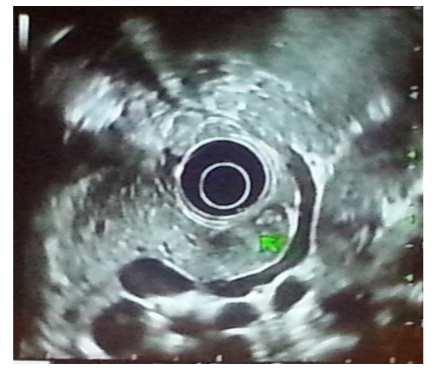
Endoscopic Ultrasound (EUS) performed showed a well-encapsulated mass of pancreatic tail measuring 22 mm in diameter, heterogeneous with some calcifications inside (Figure 1) suggesting either a neuro endocrine tumor or a pseudo-papillary tumor of the pancreas. The patient was operated and had a caudal pancreatectomy by laparoscopy without postoperative complications. The histopath-ological examination of the surgical specimen with complementary immunehisto -chemistry diagnosed (SPPT). Currently, the patient is very well.
2. Discussion
Described in 1959 by Frantz [1], the pseudopapillary tumor of the pancreas is known by various names: Frantz tumor, the papillary cystic and solid tumor of the pancreas, papillary epithelial neoplasm, Papillary cystic tumor of the pancreas. In 1996, it was classified by WHO as a tumor of the exocrine pancreas under the name of solid-pseudopapillary tumor of the pancreas (SPPT) [2]. It represents 1 to 2% of the total exocrine tumors of the pancreas [3]. It is a non- secreting tumor, which occurs mainly in young women, with an average age of 28 years and a sex ratio of 10: 1 [4]. In men, the average age of diagnosis is higher (37 years) [5].It is a tumor with a low degree of malignancy, Kato et al showed that its growth rate is slow with an estimated doubling time of 765 days [6]. Degenerative cases have been reported in approximately 15% of adult cases, particularly in elderly or male patients [7].Clinically, in more than 50% of cases the tumor is asymptomatic [8].The diagnosis is suspected by Imaging (CT, MRI, EUS): This tumor has a diameter varying between 5 and 10 cm, it can affect the head, body or tail of the pancreas, with a predominance in the Caudal region (40% of the cases). (SPPT ) is often encapsulated with a mixed tissue and cystic composition. Peripheral calcifications may exist [9,10]. The diagnosis is confirmed by the immunohisto - chemistry after surgical excision. Differential diagnosis is mainly with neuroendocrine tumors of the pancreas and mucinous cystadeno carcinomas. The biopsy should be avoided because of the risk of extra pancreatic dissemination, and the transformation of a well localized tumor with good prognosisinto an aggressive tumor, so surgery is indicated for diagnostic and Therapeutic purposes [11]. The etiopathogenesis of this tumor remained unknown until now. Several hypothesis have been reported: the feminine predominance , the presence of hormonal receptors on the surface of tumor cells has raised the hypothesis of hormone dependent [12]; On the other hand, extra pancreatic localizations have been reported, which allowed us to suggest other hypothesis, either a development from an ectopic pancreatic parenchyma, or a development from undifferentiated tot potent stem cells that migrate and differentiate secondarily into pancreatic cells[13,14].hypothesis of ductual, mucinous, neuro secretory origin were mentioned [14]. The occurrence of solid tumors in patients under immune suppressanth as been reported [15] but the use of immunosuppressant was not necessary in our case. The association of (SPPT ) with ulcerative colitis has never been reported. Is it an incidental association or is there a common point between the 2 pathologies? The most likely pathogenesis of UC, is the activation of the intestinal immune system due to one or more environmental factors to individuals with genetic suscept -ibility. In front of this association, we supposed that there may be something in common between the two diseases and 3 other setiopathogenic hypotheses can be suggested: most likely is a genetic predisposition to the tumor as the patient has already a family history of Inflammatory bowel disease (IBD), an immunological anomaly or an environmental factor. The treatment is exclusively surgical. The place of the neo-adjuvant treatments is not defined. The prognosis of solid and pseudo-papillary tumors is good. Long-term survival after complete resection of a non-metastatic tumor is excellent, ranging from 80 to 90% [16]. Survival up to 17 years for metastatic tumors has been reported[17].Long-term follow-up of these patients is necessary and particularly in men. Recurrence can occur even years after initial surgery. The mortality rate is estimated at 2% [18].
3. Conclusion
The papillary and cystic tumor of the pancreas is a rare pancreatic tumor with a low degree of malignancy. The diagnosis is confirmed by the immunehisto chemistry after surgical excision, its prognosis is excellent. The association with UC has never been reported, this suggests other pathogenic hypothesis that have to be confirmed or excluded.
References
- Frantz VK. Tumors of the pancreas. In: Atlas of Twnor Pathology, section VII. fascicles 27, 28. Washington DC: Armed Forces Institute of Pathology; 1959.
- Klöppel G. SE, Longnecker D.S., Capella C.,Sobin L.H. World Health Organisation International Histologic Classification of tumors 2. Histologic typing of tumor of the exocrine pancreas. In. Berlin: Springer-Verlag, 1996.
- Hassan I, Celik I, Nies C, et al. Successful treatment of solid pseudo papillary tumor of the pancreas with multiple liver metastases. Pancreatology 2005 ; 5 : 289-94.
- Yu PF, Hu ZH, Wang XB, Guo JM, Cheng XD, Zhang YL et al. Solid pseudo papillary tumor of the pancreas: a review of 553 cases in Chinese literature. World J Gastroenterol. 2010 Mar 14; 16(10): 1209-14.
- Machado MC, Machado MA, Bacchella T, Jukemura J, AlmeidaJL, Cunha JE. Solid pseudopapillary neoplasm of the pancreas: distinct patterns of onset, diagnosis, and prognosis for maleversus female patients. Surgery 2008;143:29—34.
- Kato T, Egawa N, Kamisura T, Tu Y, Sanaka M, Sakaki N. A case of solid pseudopapillary neoplasm of the pancreas and tumor doubling time. Pancreatology 2002 ; 2 : 495-8.
- Lam KY, Lo CY, Fan ST. Pancreatic Solid-cystic-papillary Tumor: Clinicopathologic features in height Patients from Hong Kong and Review of the Literature. World J Surg 1999; 23:1045–50.
- RomicsJr L, Oláh A, Belágyi T, Hajdú N,Gy˝ur˝us P, RuszinkóV. Solid pseudopapillary neoplasm of the pancreas-proposedalgorithms for diagnosis and surgical treatment. LangenbecksArchSurg 2010;395:747—55.
- Petrella T, Rat P, Lizard G, Dusserre-Guion L, Poulard G, Michiels R. Tumeur papillaire et kystique du pancréas. Étude histologique, immuno-histochimique et par cytométrie en flux. Gastroenterol Clin Biol 1994 ; 18 : 1021-7.
- DeestG , Gauss X , kerdaron R , piquard A Lagasse JP. Apport de l'echoendoscopie pour le diagnostic des TPPS du pancreas. Gastroenterol Clin Biol. 2008 Oct;32(10):813-5.
- Levy P, Hammel P, Rusziewski P. Ne pas biopsier les tumeurs pseudo-papillaires et solides du pancréas ! Gastroenterol Clin Biol 2009;33:501—2.
- Malluz A, Bustamante F, Silva JA, Torres Vargas S. Preoperative
- gemcitabine for unresectable, solid pseudopapillary tumor of the pancreas. Lancet Oncol 2005;6:185-186.
- Notohara K, Hamazaki S, Tsukamaya C, Nakamoto S, Kawabata K,
- Mizobuchi K et al. Solid pseudo-papillary tumor of the pancreas.Am J SurgPathol 2000;24:1361-1371.
- J. Podevin et al. . Tumeurs pseudopapillaires et solides du pancréas : à propos de cinq cas et revue de la littérature. Annales de chirurgie 128 (2003) 543–548.
- Beaugerie L CF, BouvierAM. The use of immunomodulators and biologics in inflammatory bowel diseases a cross-sectional French nationwide cohort 2004-2005. Gastroenterology 2006 130 A648-A649.
- Tait N, Greenberg ML, Richardson AJ, Osborn RA, Little JM. Frantz’s tumour: papillary and cystic carcinoma of the pancreas. Aust NZ J Surg 1995;65:237–41.
- Papavramidis T, Papavramidis S. Solid pseudopapillary tumors of the pancreas: Review of 718 patients reported in English literature. J Am CollSurg 2005;200:965-972.
- Machado MC, Machado MA, Bacchella T, Jukemura J, AlmeidaJL, Cunha JE. Solid pseudopapillary neoplasm of the pancreas: distinct patterns of onset, diagnosis, and prognosis for maleversus female patients. Surgery 2008;143:29—34.