


|
|
DOI Prefix 10.20431 |
Information
Journal Policies
Neonatal Central White Matter can Stand Short Period of Oxygen and Glucose Deprivation
Tahani R. Huria1*, Samir Elmrghni1
Copyright :© 2018 Authors. This is an open-access article distributed under the terms of the Creative Commons Attribution License, which permits unrestricted use, distribution, and reproduction in any medium, provided the original author and source are credited.
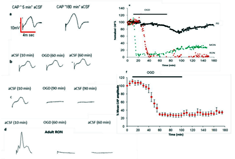
In previous studies, anoxia was used as the insult to investigate the mechanism of white matter injury in the optic nerves. Recently, ischaemia (i.e. oxygen and glucose deprivation (OGD)) has been recognised as being more clinically relevant than anoxia. As well as the ideal model to investigate the function of central nervous system and the mechanism of injury is rodent optic nerves (RONs). They have complete white matter tracts, and they are devoid of the complications of neuronal cell bodies. Under control conditions the functions of rodent optic nerves were investigated by electrophysiological recording of compound action potential (CAP). Axon function (measured as the area under the CAP) remained relatively stable for more than 120 min in both adult rat optic nerves (RONs) and mouse optic nerves (MONs) at 37°C (115.10 ± 10.5% of the initial value, (n=8) & 100.0 ± 5.5% of the initial value, (n=18), respectively. The CAPs of RONs and MONs have three discreet peaks.
We conclude that, optic nerves of developed brains are less resistant to shorter periods of oxygen and glucose deprivation (OGD) than developing optic nerves brains.
Keywords: optic nerves, action potential and ischaemia ,Forensic Science
1.Introduction
Cerebral white matter injury in the premature infant is a problem of enormous importance. For example, in the USA each year approximately 60 000 infants (1.5% of the 4000000 yearly live births) are born with a birth weight less than 1500 g,[1] and based on MRI data at least 50% exhibit some degree of cerebral white matter injury,[2,3] as defined later. This injury likely accounts for the predominance of neurological deficits observed in the approximately 90% of infants who survive. These deficits in survivors include cerebral palsy in 5–10% and importantly, cognitive/ behavioural /attentional deficits in about 50%[4,5]
Although other pathologies occur in premature infants—for example, severe intraventricular haemorrhage, periventricular haemorrhagic infarction, hydrocephalus, cerebellar disease— cerebral white matter injury seems to be the predominant lesion. Prevention of this injury requires insight into pathogenesis, and recent research holds promise that preventive interventions will be found.
2. Methodology
The compound action potentials were recorded from both neonatal rat optic nerves and adult rodent optic nerves by using the electrophysiological techniques. The functions of rat optic nerves were tested under both normal conditions and the insult of ischaemia. In normal conditions, the artificial cerebrospinal fluid was used as normal environment; it's a mixture of both 95% O2 & 5% CO2. The nerves were exposed to 10-15 min normal conditions, after that ischaemia is induced by exposing the nerves to a mixture of 95%N2 and 5% CO2 for 60 min duration. The same experiment repeated for 90 min duration.
3. Results
The standard 60-min period of OGD produced injury in adult RONs and MONs and failed to result in a significant permanent loss of the CAPs in neonatal nerves. Acute exposure to a 60-min period of OGD in day 0 and day 4 rat optic nerves (P0-P4 RONs) produced very mild and transient changes in the CAP amplitude, followed by complete recovery after restoration of normal conditions to 99. 7 ± 5.87% of the initial value, (n=8). The recovery in the CAPs recorded from P0-P4 RONs are compared to the recovery in the CAPs recorded from adult RONs and MONs after exposure to 60 min OGD, 99. 7 ± 5.8% of the initial value, (n=8); 15.2 ± 15.5% of the initial value, (n=8); and 29.3 ± 4.5% of the initial value, (n=8), respectively. These results show that, developing optic nerves are more resistant to shorter periods of OGD than optic nerves of developed brains.
References
- Hamilton BE, Minina AM, Martin JA, et al. Annual summary of vital statistics: 2005. Pediatrics. 2007; 119:345–60.
- Volpe JJ. Cerebral white matter injury of the premature infant—more common than you think. Pediatrics. 2003; 112:176–9.
- Dyet LE, Kennea N, Counsell SJ, et al. Natural history of brain lesions in extremely preterm infants studied with serial magnetic resonance imaging from birth and neurodevelopment assessment. Pediatrics. 2006; 118:536–48.
- Hack M, Taylor HG, Drotar D, et al. Poor predictive validity of the Bayley Scales of Infant Development for cognitive function of extremely low birth weight children at school age. Pediatrics. 2005; 116:333–41.
- Wilson-Costello D, Friedman H, Minich N, et al. Improved neuro developmental outcomes for extremely low birth weight infants in 2000– 2002. Pediatrics. 2007; 119:37–45.