


|
|
DOI Prefix 10.20431 |
Information
Journal Policies
Mouth Breathing-A Harmful Habit in a Young Child
Nilufer Nadaf1, Krishnapriya .V2, Shilpa G3, Santosh Challa3, Ramakrishna V.V.V4, Mayuri Ganesh4
2.Professor and Head of Department, Department of Pedodontics and Preventive Dentistry, Army College of Dental Sciences, India
3.Reader, Department of Pedodontics and Preventive Dentistry, Army College of Dental Sciences, India
4.Senior Lecturer, Department of Pedodontics and Preventive Dentistry, Army College of Dental Sciences, India
Copyright :© 2018 Authors. This is an open-access article distributed under the terms of the Creative Commons Attribution License, which permits unrestricted use, distribution, and reproduction in any medium, provided the original author and source are credited.
Mouth breathing is a habit that may develop due to various reasons. As it may lead to many harmful conditions, early diagnosis is key to treatment. A pediatric dentist may be one of the first healthcare professionals to come in contact with a patient who exhibits mouth breathing and thus it is important to have a sound knowledge to perform correct diagnosis and effective treatment.
Keywords: Mouth-Breathing, Deleterious habit, Diagnosis,Forensic Science
1.Introduction
Mouth breathing refers to the state of inhaling and exhaling through the mouth. The literature describes the prevalence of mouth breathing as ranging from 5 to 75% of tested children[1].The mouth does not usually contribute in respiration. Increased struggle to the flow of air through the nasal passage may be considered to be the key reason of mouth breathing[2].
Nasal obstruction forces normal nasal breathing into oral breathing[3]. Sassouni determined mouth breathing as a habitual respiration through the mouth instead of the nose[4]. Mouth-breathing (MB) is a substitute respiratory mode and consists of a mechanically incorrect form of respiration. It has a multifactor etiology and multiple consequences[5].
Mouth breathing is a deleterious habit.4Due to its range of co-morbidities, mouth breathing (MB) has been a concern for healthcare professionals in various areas. Mouth breathing (MB) is also an etiological factor for sleepdisordered breathing (SDB) during childhood. Both habit and obstruction may cause facial muscle imbalance and craniofacial changes.
MB is one of the most commonly cited characteristics of sleep-disordered breathing (SDB) during childhood, but symptoms are often inadequately recognized. SDB encompasses a wide clinical spectrum, such as snoring, upper airway resistance syndrome (UARS), and obstructive sleep apnea (OSA). Dentists may be the first healthcare professionals to have contact with a MB child[6].
2. Classification
Slim and Finn classified mouth breathing in 1987 as:
a) Obstructive
b) Anatomical
c) Habitual[4]
The purpose of breathing is to oxygenate the body and to remove the waste carbon dioxide[4]. The nose and the mouth have different functions. Each nostril functions independently and synergistically to filter, warm, moisturize, dehumidify and smell the air[7] Nose breathing controls the volume of inhaled and, more important, the volume of exhaled air.
Body oxygenation occurs during exhalation not during inhalation[4]. Breathing is subconscious with each inhale determined not by the need for oxygen, but by the level of carbon dioxide in the lungs and blood. The back-pressure created in the lungs with the slower exhale of nose breathing compared to mouth breathing allows more time for the lungs to transfer oxygen to the blood. Thus, nasal breathing will increase oxygen in the lungs, blood and cells[7].
Mouth breathing to take in more air does not increase the level of oxygen in the blood, which is already 97-98 percent saturated. Mouth breathing with big breaths actually lowers the carbon dioxide level in the lungs and the blood leading to lower levels of oxygen released from the haemoglobin to the body cells. Excessive carbon dioxide loss through mouth breathing decreases oxygen in the lungs, blood and cells[7]
The etiology of mouth breathing is multifactorial. Mouth breathing is majorly caused due to nasal obstruction. Nasal obstruction can result from either congenital or postnatal causes and may amplify resistance to air-flow and impair sucking-swallowing responses, with increased risks of aspiration or of more severe and threatening respiratory distress conditions. In addition, nasal obstruction alters the “trophic” flow of sensory information towards the olfactory brain[3].
The most common cause of MB is the presence of obstacles in the nasopharyngeal region, which increases nasal resistance. The most extreme forms are due to congenital laryngomalacia, bilateral choanal atresia, or oronasal defects associated with Pierre Robin syndrome. Less extreme forms involve choanal stenosis, unilateral choanal atresia, or defects of the nasal septum related to cleft palate. Other mechanical causes such as those due to obstructive tissue masses (adenoid or/and tonsillar hypertrophy) prevail during later development. More benign, short-term obstructive forms derive from mucosal accumulation due to neonatal infections or allergic rhinitis[3]. Intranasal defects such as deviated nasal septum, bony spurs or polyps may also cause mouth breathing.
An early diagnosis is crucial for the correction of mouth breathing and to avoid any associated conditions. Correctly diagnosing a habit of mouth breathing may require a detailed case history, clinical examination and diagnostic tests.
A detailed case history regarding the development of the habit, duration, frequency and associated symptoms must be recorded. This should be followed by a clinical examination of the intraoral and extraoral features of the patient and making a record of the same.
It is important to fully diagnose a patient with the habit of mouth breathing through various clinical tests available. The mirror test and the water retention test are among the breathing tests most cited in the literature. The lip seal test was the most frequently used, followed by the mirror test and the water retention test[6].
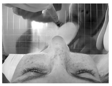
The mirror test is also called the fog test. A double-sided mirror is held between the nose and the mouth. Fogging on the nasal side of the mirror indicates nasal breathing while fogging on the oral side-mouth breathing. (Figure 1).
Massler and zwemer butterfly test/cotton test-Butterfly shaped cotton strands are placed over the upper lip below nostrils. On exhalation if the fibres flutter downwards, the patient is a nasal breather and if the fibres flutter upwards, the patient is a mouth breather.
Inductive plethysmography (rhinometry): The total airflow through the nose and the mouth can be quantified using inductive plethysmography. This allows the percentage of nasal and oral respirations to be calculated.
Cephalometrics: It can be used to calculate the amount of pharyngeal space, size of adenoids and to know the skeletal pattern of the patient[2].
3. Mechanism
The change in the way of breathing leads to a change in the jaw, tongue and head position. The balance between the tongue action, on the one hand, and the mimic and masticatory muscles, on the other hand, is disturbed. The “forming” action to the mid-face of the air passing through nasal cavity is disturbed too, and it influences normal palatal development. In mouth breathing pattern the tongue is usually shifted back and downwards and doesn’t participate in the development of the hard palate, which results in the formation of a deep gothic palate. A forward head posture is developed in order to make easier inhalation through mouth, the lower jaw is underdeveloped and placed downward and backwards, and this leads to its distal position and over jet formation. Taut cheek muscles apply an increased external force to the upper jaw which causes V shaped form.6 The low and forward position of the tongue is common in the presence of hypertrophic palatine tonsils as an attempt to increase posterior airway space and ease breathing causes the Mandible to drop, causing an Imbalance between masticatory, mimic and tongue muscles leading to Adenoid facies or long face syndrome.
4. Treatment
According to symptoms:
1. Application of petroleum jelly on the gingiva.
2. Remove the cause: Etiological factors should be treated first. Removal of nasal and pharyngeal obstruction should be sought. If a respiratory allergy is present, it should be brought under control.
3. Intercept the habit: If the habit continues after the removal of the obstruction, it should be corrected.
a. Hold a pencil between the lips.
b. Hold a sheet of paper between the lips.
c. Button pull exercise-A button of 1 and a half diametre is taken and a thread is passed through the button hold. The patient is asked to place the button behind the lop and pull the thread, while restricting it from being pulled out by using lip pressure.
d. Tug of war exercise-this involves 2 buttons, with one placed behind the lips while the other button is held by another peson to pull the thread.
Advocated by Macaray in 1960, these expanding exercises are used in conjunction with the macaray activator. It is constructed with aluminium with which the development of dental arches and dental base relationship could be corrected at the same time.
First introduced by Newell in 1912.It is a myofunctional appliance that is easy to fabricate and easy to wear. It works on the principle of both force application and force elimination.2
A new treatment protocol given by Denotti et al in 2014 includes a rapid palatal expander bonded on the decidous molars, in association with the use of silicone myofunctional devices such as the nasal stimulator and the oral obturator, that speeds and increases the effectiveness and stability of treatment[8].
5. Effects
When abnormal pressure of muscles interferes in facial growth, it can determine the appearance of a malocclusion. Unbalanced facial musculature occurs as a result of MB, which causes changes in tooth positioning, lips, tongue, palate, and jaws, so as to counterbalance the new breathing pattern. The most prevalent malocclusions found in the mouth-breather group were atresic palate and anterior open bite[6].
6. Features
Mouth breathing patients have characteristic intraoral and extraoral symptoms. In the period between 1970 and 1980, Linder-Aronson reported the connection between mouth breathing and craniofacial appearance including long face, anterior open bite, overjet, posterior crossbite[4].
Following extraoral signs are apparent:
a. lip incompetence
b. a short upper lip
c. dry and cracked lips
d. an increased lower facial third;
e. an increased mandibular angle
f. dark circles under the eyes
g. narrow nostrils
h. a small and tip-tilted nose
i. smoothed nasolabial folds
j. A typical head position – in extension
k. The most common and characteristic intraoral signs include
l. a deep or gothic palate
m. a V-shaped upper dental arch
n. posterior crossbite
o. anterior open bite
p. an overjet
q. class II malocclusion
r. gingivitis in frontal teeth[4]
Short attention span, decreased memory, poor school performance, changes in sleep-wake cycle, day irritability and then a negative influence on psycho-social life of the subject are all aspects related to a poor oxygenation[8].
7. Discussion
General and pediatric dentists may be in the best position to screen and treat patients who suffer from upper airway obstruction/mouth breathing. Dentists usually see patients on a regular basis every six months, and swollen tonsils can be easily detected by using a mouth mirror to look at the back of the patient’s throat. All patients— children, adolescents, and adults—should be screened for upper airway obstruction. Mouth breathing (and all of its associated medical, social, and behavioural problems) is best managed by using a multidisciplinary approach involving pediatricians, physicians, dentists, and ear-nose-throat (ENT) specialists[9].
Malhotra S et al found out that children who breathe predominantly through their mouth pose difficult problems for healthcare professionals. It was also found that all subjects with mouth-breathing habit exhibited significant increase in facial height, mandibular plane angle and gonial angle[10].Conti et al observed that Postural problems were significantly more common among children in the group with mouth breathing syndrome, highlighting the need for early interdisciplinary treatment of this syndrome[11]. Nunes et al discovered that Different sites of obstruction of the upper airway due to enlarged lymphoid tissue are associated with different types of dental malocclusion. Findings are relevant to orthodontic and surgical decision making in these mouth-breathing patients[12]. Udaka et al observed that chronic nasal obstruction impairs Quality of Life, at least partially, through excessive daytime sleepiness possibly caused by sleep‐disordered breathing[13]. Souki et al observed that the prevalence of posterior crossbite is higher in mouth breathing children than in the general population. During mixed and permanent dentitions, anterior open bite and class II malocclusion were more likely to be present in mouth breathers[14]. Gulati et al observed that the Gingival index was found to be higher in the mouth breathers than in the normal breathers in the subjects with incompetent lip seal. Increased lip separation and decreased upper lip coverage were all associated with higher levels of Plaque index and Gingival index. No statistical difference existed between mouth breathers and normal breathers with respect to Plaque index[15].
References
- Triana BE, Ali AH, León IB. Mouth breathing and its relationship to some oral and medical conditions: physiopathological mechanisms involved. Rev haban cienc méd. 2016; 15(2).
- Jain A, Bhaskar DJ, Gupta D, Yadav P, Dalai DR, Jhingala V, Garg Y, Kalra Mouth Breathing: A Menace to developing dentition. J Contemp Dent 2014; 4(3): 145-151.
- Trabalon M, Schaal B. It takes a mouth to eat and a nose to breathe: abnormal oral respiration affects neonates' oral competence and systemic adaptation. International journal of pediatrics. 2012 Jul 3; 2012.
- Valcheva Z, Arnautska H, Dimova M, Ivanova G, Atanasova I. The role of mouth breathing on Dentition development and formation. Journal of IMAB 2018 Jan-Mar; 24(1): 1878-1882.
- Milanesi JD, Weber P, Berwig LC, Ritzel RA, Silva AM, Corrêa EC. Childhood mouth-breathing consequences at adult age: ventilatory function and quality of life. Fisioterapia em movimento. 2014 jun; 27(2): 211-8.
- Pacheco MCT, Casagrande CF, Teixeira LP, Finck NS, Araújo MTM. Guidelines proposal for clinical recognition of mouth breathing children. Dental press j orthod. 2015 july-aug; 20(4): 39-44.
- O’Hehir T, Francis A. Mouth Vs Nasal Breathing. Hygiene and prevention. September 2012: 120-123.
- Denotti G, Ventura S, Arena O, Fortini A. Oral breathing: new early treatment protocol. J Pediatr Neonat Individual Med.2014; 3(1): e03 0108.
- Jefferson Y. Mouth breathing: adverse effects on facial growth, health, academics, and behavior. Gen Dent. 2010 Jan; 58(1): 18-25.
- Malhotra S, Pandey RK, Nagar A, Agarwal SP, Gupta VK. The effect of mouth breathing on dentofacial morphology of growing child. Journal of Indian Society of Pedodontics and Preventive Dentistry. 2012 Jan 1; 30(1): 27.
- Conti PB, Sakano E, Ribeiro MÂ, Schivinski CI, Ribeiro JD. Assessment of the body posture of mouth-breathing children and adolescents. Jornal de pediatria. 2011 Aug; 87(4): 357-63.
- Nunes WR, Di Francesco RC. Variation of patterns of malocclusion by site of pharyngeal obstruction in children. Archives of Otolaryngology–Head & Neck Surgery. 2010 Nov 15; 136(11): 1116-20
- Udaka T, Suzuki H, Kitamura T, Shiomori T, Hiraki N, Fujimura T, Ueda N. Relationships among nasal obstruction, daytime sleepiness, and quality of life. The Laryngoscope. 2006 Dec; 116(12): 2129-32.
- Souki BQ, Pimenta GB, Souki MQ, Franco LP, Becker HM, Pinto JA. Prevalence of malocclusion among mouth breathing children: do expectations meet reality?. International journal of pediatric otorhinolaryngology. 2009 May 1; 73(5): 767-73.
- Gulati MS, Grewal N, Kaur A. A comparative study of effects of mouth breathing and normal breathing on gingival health in children. Journal of the Indian Society of Pedodontics and Preventive Dentistry. 1998 Sep; 16(3): 72-83.