


|
|
DOI Prefix 10.20431 |
Information
Journal Policies
The Importance of Panoramic Radiography as an Auxiliary Instrument in Clinical and Legal Dental Practices
Barbieri, Ana Amélia1,5,Almeida, Renata Costa2, Naressi, Suely Carvalho Mutti3, Moraes, Luiz Cesar4, Moraes, Mari Elí Leonelli4
2.DDS, São Paulo Dentistry Association -APCD Section of São José dos Campos, São Paulo, Brazil.
3.DDS, MSc, PhD, Associate Professor, Department of Social Dentistry School of Dentistry of São José dos Campos, São Paulo State University-UNESP,Brazil.
4.DDS, MSc, PhD, Associate Professor, Department of Biosciences and Oral Diagnosis School of Dentistry of São José dos Campos, São Paulo State University-UNESP,Brazil.
5.Rua Conselheiro Moreira de Barros 56 apto 94 - Centro, Taubaté – São Paulo,CEP,Brazil.
Copyright : © 2016 Barbieri AA. This is an open access article distributed under the Creative Commons Attribution License, which permits unrestricted use, distribution, and reproduction in any medium, provided the original work is properly cited.
Abstract
The aim: This study was conducted in order to found the inclusion of panoramic radiography as a vital document in the patient's medical history, giving it more reliability. Methods: For this purpose, 2,732 un-identified panoramic radiographs were evaluated, from patients aged from 15 to 55, males and females, from the files of the Radiology Department from the Dental School of São José dos Campos - São Paulo State University, and from a private radiology clinic in the city of São José dos Campos - SP. During analysis of the radiographs, non-clinically observable radiographic findings were sought. The data found were submitted to descriptive statistical analysis and to the Student T-test. Results: Of all the panoramic radiographs evaluated, 20.31% presented radiographic findings capable of interfering with the diagnosis and prognosis most appropriate for the health of the individual. No statistically significant differences were found between the genders or between gender and age bracket. Conclusion: The results obtained and the multiple odontological purposes of panoramic radiography demonstrate its importance as a document to supplement the odontological history of the individual, giving it more legitimacy and clinical value.
Keywords: Panoramic Radiography, Documentation, Forensic Dentistry, Diagnosis.
1.Introduction
Dental clinical practice is not based simply on clinical care, but also on the relationship between the patient and the dental professional. When seeking dental care, the individual makes sure that the professional who will perform the treatment is reliable, and places his chances of success in the treatment on their interpersonal relationship and on the history of the professional. The relationship established based on their mutual interests is understood by current legislation as a commercial relationship within which services are rendered, even if there is no formal documentation such as a contract for dental services rendered. This happens because, under the law, the contract for the rendering of services can be carried out only by the declaration of presumed will (tacit contract). Thus, under no circumstances may dental surgeons be exempted from the responsibility that may occasionally fall on them due to procedures or treatments performed under the allegation that a legal contract was not formalized.
Professional accountability is characterized by the non-fulfillment of an obligation by the agent but may be counteracted by the patient's negligence. The transitory legal relationship established between agent and patient, whose object consists of a service rendered between them, is called an obligation.
Due to the ethical and legal aspects that envelop the patient/dental professional relationship, which it is the dental surgeon's duty to know, the Brazilian Dental Ethics Code discusses the dental patient's history (physical and/or electronic files of all the documents related to the treatment)[2]. In this publication the patient's history was divided in the following manner: fundamental documents: clinical file containing the identification of the dental professional and patient and the clinical examination records, anamnesis, treatment plan, evolution and incidents, prescriptions, certificates, contract for dental services rendered, and supplementary exams, as well as the radiographic exams; supplementary documents: any documents related to the treatment, as post-surgical recommendations. All the documents contained in the patient's history, individually or together, may be submitted to the analysis of the technical assistants and/or legal experts to clarify questions stemming from legal disputes about the procedures performed by the dental surgeon on a specific patient. These documents will help the technical assistants in the preparation of their inquiry and they will allow the legal expert to make an accurate evaluation of previous procedures and of the treatment proposed and performed. The radiographic examinations, capable of recording the before and after, play an important role on these occasions. The patient's dental history does not have only a contentious purpose. On the contrary, it has a very important role in the process of human identification, for existing documents such as radiographs in the patient's dental history combine records of unique, immutable, and perennial characteristics of the individual, and therefore have the biological requirements fundamental to the process of human identification: uniqueness, individuality, immutability, and permanence. The correct and thorough completion of a patient's history facilitates and accelerates the tasks of the legal expert in the field. Positive identification or the exclusion of identity may be obtained by the comparison of an individual's characteristics and specific parameters recorded in the present with records made previously. Radiographic images maintain the record of countless and different findings capable of contributing to the identification process of an individual. In addition, through radiographic analysis, it is also possible to perform a chronological examination of eruption and crown-root mineralization, which can provide a more precise age estimate. Among the existing methods for the estimation of age, the dental method comes the closest to the real age of the individual. As we can see, radiographic examinations have multiple legal dental purposes in addition to other essentially clinical.
Radiographs are the supplementary examinations most frequently performed by dental surgeons. Panoramic radiography is the broadest among dental radiographs today. It provides a complete view of the facial structures and, clinically, it helps in the diagnosis and in the therapeutic planning for treatment of diseases involving teeth and facial bones, revealing findings that the clinical exam would not detect and that could compromise the prognosis of a treatment. In addition to the clinical benefits already described, the inclusion of this radiography in the patient's history will propitiate quicker and more impartial solutions to legal questions pertaining to civil, penal, and ethical-administrative lawsuits, and would help in the forensic processes of identification and age estimation.
The clinical and legal benefits that can be had with the utilization of panoramic radiography motivated this study, whose objective was to promote this type of radiography as an auxiliary instrument in the diagnosis at the beginning of treatment and as a legal dental document supplementing the patient's dental history.
2.Material and Methods
The research was approved by the Research Ethics Committee from the Dental School of São José dos Campos - São Paulo State University under protocol no. 076/2009.
The sample, representative of the population studied, was by interest and non-probability. It used 2.732 digital panoramic radiographs of individuals aged between 15 and 55, males and females, obtained in the period between January and June of 2010. Of these 2.732 digital panoramic radiographs, 615 came from the files of the Radiology Department of the Dental School of São José dos Campos - São Paulo State University (Dental X-ray machine ORTHOPHOS XG 5 DS manufactured by Sirona Dental Systems - Bensheim, Germany) and 2.117 from a private radiological clinic in the city of São José dos Campos - SP (Dental panoramic X-ray machine Kodak 9000 3D manufactured by Kodak Trophy - Marne La Vallée, France).
The researchers did the survey, analysis, and the notations of all the data for the study. Gender, age, and the radiographic findings were noted and given identifying codes (Chart 1).
The images suggestive of abnormalities utilized in this study followed the current International Classification of Diseases to Dentistry and Stomatology (ICD-DA), considering the radiographically visible alterations, that is, of hard tissue. The condyloid alterations, such as osteophytes, were all combined into one category. Unerupted and impacted teeth were considered for the effect of this study, as were retained teeth. The criterium to consider an element as retained was that the element had two thirds or more of its root formed and had not erupted.
The images of clinically observable alterations and those referring to symptomatic pathological processes were not considered in this study, for their reports call for supplementary examination. In the same way, images of dental treatments such as teeth treated endodontically, pins for implants, and fixed prostheses, among others were not considered.
The data were submitted to descriptive statistical analysis and the Student T-test for equivalent variables. The calculation of the statistical differences was made considering the percentage by gender of the total sampling, compared separately for anomalies and images suggestive of pathological processes.
3.Results
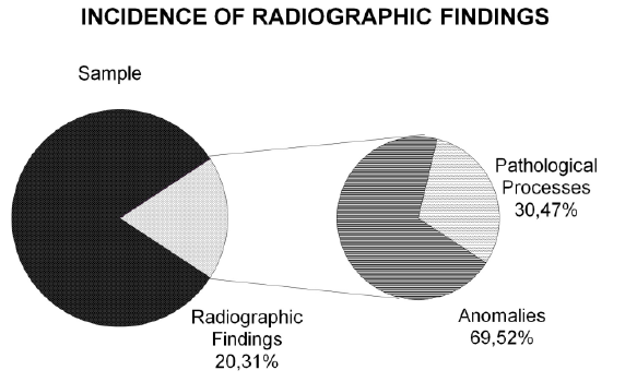
From the total of radiographs analyzed, 1,214 were from males (44%) and 1,518 were from females (56%). Radiographic findings were observed in 555 radiographs (20.31% of the sample), and were evaluated statistically considering the dental anomalies and the pathological processes separately. (Figure 1)
From the sample analyzed, 431 panoramic radiographs showed some anomaly. There was the simultaneous occurrence of 2 anomalies in 64 radiographs and of 3 anomalies in 2 radiographs. According to the T-test for average, considering equivalent variables and the analyses that show anomalies, there was no difference of age statistically significant for either gender. (Table 1) The number of anomalous occurrences in males and females did not differ significantly from the proportion of the sample, P: 1.25.
Of the anomalies found, 99% corresponded to retained teeth, dilaceration, supernumerary, micro teeth, and agenesis.The most prevalent anomaly was retained teeth (64% of the cases), followed by dilaceration (14% of the cases) and supernumerary (13% of the cases). Together these anomalies accounted for more than 90% of the cases (Figure 2).
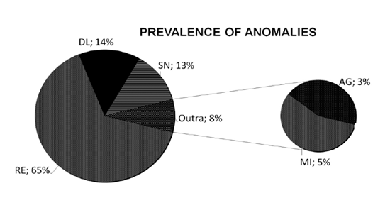
RE: Retained teeth DL: Dilaceration SN: Supernumerary AG. Agenesis MI: Microtooth
When specifically verifying the occurrence of each anomaly in relation to gender, no statistically significant difference was found, considering a significance of 1%. (Table 2).
From the sample analyzed, 188 panoramic radiographs have shown some image suggestive of pathological processes, and 12 of them more than one.
According to the T-test for average, considering equivalent variables, there was no statistically significant difference in age for the genders between the radiographs that have shown images suggesting pathological processes. (Table 3)
The number of occurrences of pathological processes in males and females did not differ significantly from the proportion of the sample, P: 1.25. The prevalent pathological processes were condyloid alterations (47%) and alterations in the styloid process (29%). Together these pathological processes made up more than 50% of the cases. Combined with the odontoma and sialolithiasis they represent 87% of the pathological processes found. (Figure 3)
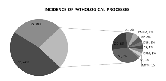
CD: Condyloid alteration ES: Styloid process alteration SL: Sialolite OD: Odontoma CG: Dentigerous cyst CMSM: Maxillary sinus mucous cyst DP: Periapical dysplasia CNP: Nasopalatine cyst CS: Stafne´s Cyst DFM: Bone defect MTM: Soft tissue mineralization AR. Atherosclerosis.
Analyzing the occurrence of pathological processes specifically related to gender, no significant statistical differences were observed, considering a significance of 1%. (Table 4)
4. Discussion
The radiographic findings shown in this research were sub-divided into images suggestive of pathological processes and anomalies, to provide a broad view of the possibilities of radiographic findings while they are also grouped by their characteristics, particularities, and possible clinical consequences. Of the 2,732 radiographs evaluated, 20.31% showed radiographic findings. (Figure 2) This result coincides with the findings by Cholitgul and Drummond[7].
Although they had methodological differences as to age and findings when compared between themselves and with this study, the studies that evaluated the radiographic findings named by their authors as "abnormalities" and "significant alterations"16,1, found equally important prevalence indices, leading the authors to conclude that it is important to detect these findings in clinical practice. The sample utilized in this study (2,732 radiographs) represents a population in the age bracket evaluated, which allows the extrapolation of the results found for the 343,151 individuals represented.
The number of occurrences of images suggesting anomalies in males and females did not differ significantly from the sample proportion (Table 2). Vicci and Capelozza[23] found a different occurrence of radiographic findings between the genders; however, they did not mention the representation of each gender in the sample. In relation to the prevalence of pathological processes corresponding to gender and age, again no statistically significant differences were observed (Table 3). Considering the age bracket, no statistically significant difference was observed among the panoramic radiographs that have shown anomalies (Table 1), corroborating what was reported by Pinheiro, et al.[19]
The anomaly most prevalent in this study was retained teeth (Table 2, Figure 2), which coincides with what was found by Keith16 and Allatar, et al.[1]. A great number of dental anomalies of form and position can be observed simply through radiographs. The early diagnosis of a retained tooth, for example, is fundamental to decide which clinical procedure is the most appropriate to be recommended by the dental professional. Certainly any non-observance of findings as prevalent as this and the absence of appropriate treatment would leave the dental professional helpless in a legal dispute.
The images suggesting pathological processes found in 188 panoramic radiographs corresponded to 6.8% of the sample. In terms of promoting health, the percentage of images suggesting pathological processes was significant, since they can result in esthetic, phonetic, and functional damages to the patient and consequently foment the culpability of the dental professional who did not observe them.
Some authors directed their survey of images suggesting pathological processes to regions and/or specific processes, such as Costa, et al.[8], who evaluated alterations in the maxillary sinus and Jacome and Abdo[15] who evaluated the calcification of soft tissues, differently from the methodology adopted in this study, which searched for images suggesting pathological processes in general.
The pathological processes prevalent in this study were condyloid alterations (47%) and alterations in the styloid process (29%) (Figure 3, Table 4). The osteophytes, degenerative condyloid alterations due to articulation problems, represented 32% of the cases in this study. Alterations in the styloid process may be related to symptoms in the region of the ear, throat, and neck. In addition to reports from individuals, a preliminary supplementary exam such as panoramic radiography is fundamental for the diagnosis, given the broadness of its image.
The results obtained have shown the clinical range of panoramic radiographic incidence and express the relevance of this auxiliary exam, able to widely record the facial structures, making it essential for an appropriate diagnosis and leading to the proposal of a treatment plan that will bring the best prognosis. The anomalies and the pathological processes not observable clinically, even with the help of intraoral radiography, but with the potential to interfere with the proposed treatment plan, can be easily seen using panoramic radiography, as reported by Monsour[18], conferring more confidence and more assertiveness to the dental professional. It is the responsibility of the dental professional to acknowledge and notify the patient or legally responsible party of any findings that may be potentially damaging to the correct development of the patient's oral health, and of the dental treatment most appropriate for their recovery, influencing the patient's general health, such as in cases of the visualization of images suggesting carotid atheroma, knowledge of which helps to prevent and provide early treatment of serious asymptomatic arteriopathies[3].
The applicability of the records contained in panoramic radiography is not limited to clinical practice, but extends to legal aspects that involve the patient-dental professional relationship and, in that aspect, is equally important and comprehensive. The records contained in it can help in analyzing the suitability of the treatment in the cases legally disputed, which nowadays is not so uncommon. The images recorded in panoramic radiography are legally conclusive and facilitate not only this process together with the other documents that compose the patient's history, but also the work of a legal expert in the study of each case, favoring an impartial decision.
Over the years, the literature has emphasized the importance of keeping all the dental documents related to patients' treatments on file. The literary reports and the need to comply with Article 5, item VIII, from the Dental Ethics Code led the CFO to request a report suggesting the classification of the documents related to dental treatments. At the time of this request, Almeida, et al.[2] proposed the following classification: fundamental documents and supplementary documents.
The CFO's proposal shows the growing concern with the creation of means that allow a better management of information and better control of professional performance. Health professionals are constantly asked to explain in detail all the procedures performed in the treatments they have given due to discussions had with patients, administratively or judicially. The administrative and legal actions are subordinate to the analysis of evidence (by witness and/or documental), affirming the concern of the CFO and the authors with the creation and maintenance of correct and complete patient history records. Nevertheless, the actions taken until the present moment in relation to this theme have not been sufficient. More effective actions are needed from the class entities in order to establish directives to standardize the documents that compose the patient's history - also because, according to the Dental Ethics Code, the non-preparation and filing of a patient's history is already an ethical infraction.
Traditionally the preparation of the documentation was designed to prove that the treatment was effectively performed and that the patient should pay for it. Currently, the documentation to be prepared must observe the possible options of treatment and the one chosen by the patient, after clarifying the risks, costs, and benefits: to present a complete anamnesis, filled in by the patient himself and directed to the treatment to be performed; to be sufficient as to requests and correct filing of various radiographic exams and other supplementary exams in order to support the planning and execution of the treatment selected; to present data and details of the evolution of the treatment in addition to reports of absences and delays, alterations in the proposed treatment due to supervenient situations, various orientations, certificates, notations of timely or late payments, referrals to specialists, medical authorizations for procedures that involve known or foreseen risks, corroborations of abandonment of treatment by the patient, and the making of any copies from the patient's history among others. This set of documents forms the dental history of each patient, and it is a defense instrument for the professional in the case of any charges of inappropriate treatment, of non-satisfaction with the result obtained, with the delinquency of professional fees, and other situations that may create a demand between the parties. The documents related to the treatment are the most effective instruments, together with the reports of the dental professionals, to permit an impartial solution for the dispute, which is increasingly common nowadays[9].
The economy, practicality, and efficiency corroborated by the utilization, separately or combined, of the documents that compose the dental patient's history are determining factors favorable to its precondition [20].
Panoramic radiography, as pointed out by Bissoli[4] and Buchner[6] was of great importance as one of the supplementary exams most requested by dental surgeons and essential to the patient's history, and which helps in the diagnosis and prognosis, in the estimation of age and in human identification. The utilization of panoramic radiography at the beginning of treatment is applicable as much in the confirmation of the appropriate treatment and recording of dental conditions before the intervention of a dental professional as in the detection of radiographic findings that would interfere with the evolution of the proposed treatment. To obtain this information and to communicate all the elements capable of interfering with the treatment to the patient are the duties of the dental surgeon, just as it is the duty of the patient to collaborate in the development of the works.
Panoramic radiographs propitiate the capturing and recording of anomalies and pathological processes present in the entire bucco-maxillo-facial structure of the individual and of all the dental procedures performed until the moment of the incidence[6]. Besides, the exposure of the patient to only one radiographic incidence satisfies the justification principle described in the Regulating Decree 453/98 from ANVISA (Portaria Normativa 453/98 from Agencia Nacional de Vigilancia Sanitaria - National Agency for Sanitary Vigilance)[5]. The radiographic dossier enormously facilitates the legal forensic expert work in resolving disputes, as already widely discussed, making it possible for the professionals involved to apprehend the clinical details that involve each case.
The legal employment of panoramic radiographs is not restricted to judicial disputes, but extends also to one of the forensic works in campus: human identification. Many dental anomalies of form and position combine the characteristics necessary for the process of human identification, and can be seen only radiographically. The range of panoramic radiographic incidence provides confidence, assertiveness, and celerity to the process of identifying bodies; facilitating the work of coroners, and diminishing the waiting time for the victims' relatives. Human identification is essential not only for humanitarian reasons, but also for helping in civil and/or criminal investigations[24], which constantly rekindles the discussion about its methods.
The utilization of patient's history documents for the identification of bodies in disasters that involve a great number of victims is more and more common, as is the number of environmental disasters and catastrophes[17]. The number of occurrences and of victims demands faster and more efficient means of identification. In this day and age, the digitization of documents that compose the dental patient's history seems to be a technologically possible solution and a front line provision, as already reported by Rothwell[21] in a literature review made to verify the importance of Odontology in human identification, in which it was concluded that the utilization of information resources in Legal Odontology saves the professional from spending countless hours comparing data ante and post-mortem in accidents with a great number of deceased victims, which increases the efficiency of the identification team. There are also efforts from the INTERPOL (International Police) in this direction: gathering professional teams from various countries to identify the victims of mass disasters[14]; developing and improving software to deal with the most varied data collected in campus; suggesting joint efforts in the standardization of dental documents to compose a patient's history, emphasizing the sufficiency of panoramic radiography in the body identification process. The evidence found between radiographs obtained ante-mortem and post-mortem is irrefutable and is therefore reliable in cases of human identification[11,13].
Equally important are the expert examinations to estimate age, for the dental estimation comes the closest to the chronological age of the individual, especially in the dental development phase[12]. These expert examinations for age estimation in the legal ambit can be a determining factor in penal imputability and/or the application of protective measures in cases of adolescents in conflict with the law[22], as well as a guarantee of civil rights.
All these things highlight the need for a new way of thinking, and for a new vision of this subject. The entities regulating the dental profession need to gather and discuss the theme effectively. New proposals are necessary for patient care, as well as the appropriate legal orientation for the protection of both patient and dental professional. It is necessary to implement a new way of storing and maintaining the documents that compose the patient's history, and streamlining the access to information. A broad discussion about the subject will benefit the Odontology professionals in all areas of this field and it will enormously facilitate the work of Legal Odontology professionals.
5.Conclusion
Panoramic radiography has multiple dental purposes and the results obtained express the relevance of this exam when considering the clinical and legal aspects that involve the relationship between patient and dental professional. Because of this, it is concluded that the inclusion of panoramic radiography as a document is vitally important to the patient's history, conferring it more legitimacy and clinical value.
References
- Allatar MM, Baughman RA, Collett WK. A survey of panoramic radiographs for evaluation of normal and pathologic findings. Oral Surg Oral Med Oral Pathol. 1980;50(5):472-8.
- Almeida CAP, Zimmermann RD, Cerveira JGV, Julivaldo FSN. Dental records - A guide to the requirement contained in item VIII, article 5 of the Dental Ethics Code. Rio de Janeiro, 2004. [access 2009 Jun 02]. Available at: http://cfo.org.br/wp-content/prontuario_2004.pdf.
- Barros RQA, Oka RCR, Holmes TSV, Cavalcanti AL, Bento PM, Godoy GP. Early diagnosis of atheroma by routine dentistry radiographic examination: recent considerations and findings. Odonto Clin Cient. 2011; 10(2):129-31.
- Bissoli CF, Castilho JCM, Medici Filho E, Moraes LC, Moraes MEL, David AF. Importance of panoramic radiography in everyday life of the dentist. Espelho. 2007; 61:9-11.
- Brazil. National Agency for Sanitary Vigilance. ANVISA Regulating Decree 453/98; 1998.
- Buchner A. The identification of human remains. Int Dent J.1985;35(4): 307-11.
- Cholitgul W, Drummond BK. Jaw and tooth abnormalities detected on panoramic radiographs in New Zealand children aged 10-15 years. N Z Dent J. 2000;96(423):10-3.
- Costa CMAC, Madeiro AT, Bandeira FG, Cunha PASMA. Diagnosis of changes in the maxillary sinuses through the scanned image. Salusvita. 2007;26(1):11-21.
- Fenelon S. Legal and ethical aspects in imaginology. Radiol Bras 2003; 36(1):III:VI.
- Glass RT. Forensic dentistry: in a terrorist world. NY State Dent J. 2005; 71(3):21-25.
- Goldstein M, Sweet DJ, Wood RE. A specimen positioning device for dental radiographic identification-image geometry considerations. J Forensic Sci. 1998;43(1):185-9.
- Goncalves ACS, Antunes JLF. Age estimation in children based upon permanent teeth mineralization degree, with a forensic dentistry scope. Odontologia e Sociedade. 1999;1(2):55-62.
- Hanaoka Y, Ueno A, Minaguchi K, Kajiwara M, Sato Y, Oshida M. Advantages of the digital x-ray system in dental identification of persons with reference to two murder cases. J Forensic Odontostomatol. 2001; 19(2):22-5.
- INTERPOL. Disaster victims identification guide - DVI Guide [Internet]. 2009; 55p. [access 2010 marco 10]. Disponivel em: http://www.interpol.int/INTERPOL-expertise/Forensics/DVI
- Jacome AMSC, Abdo EM. Radiographic aspects of soft tissues calcification in maxillofacial region. Odontol Clin-Cient. 2010;9(1): 25-32. Portuguese.
- Keith DA. The detection of abnormalities in the jaws. A survey. Br Dent J. 1973 Feb;134(4):129-35.
- Kieser JA, Laing W, Herbison P. Lessons learned from large-scale comparative dental analysis following the South Asian tsunami of 2004. J Forensic Science. 2006;51(1):109-12.
- Monsour PA. Getting the most from rotational panoramic radiographs. Aust Dent J. 2000 Jun;45(2):136-42.
- Pinheiro CC, Tostes MA, Pinheiro AR. Prevalence of dental anomalies of number in orthodontic pacients: a radiographic study. Pesq Bras Odontoped Clin Integr. 2008;8(1):47-50.
- Pretty IA. Forensic dentistry: Identification of human remains. Dent Update 2007;34:621-2, 624-6, 629-30.
- Rothwell BR. Principles of dental identification. Dent Clin North Am 2001;45:253-70.
- Silva RF, Marinho DEA, Botelho TL, Caria PHF, Bérzin F, Daruge Junior E. Determination of age by dental and wrist joint radiograph analyses: a forensic case report. Arq Odontol. 2008,44(2):93-8.
- Vicci JG, Capelozza ALA. Incidence of dental and osseous lesions to evidence through an panoramic radiographs. Rev Fac Odontol Lins. 2002;14(2):43-6.
- Warnick A. Mass disaster management: the organization of a mass disaster dental identification team. Alpha Omegan 2002;95:25-37.