


|
|
DOI Prefix 10.20431 |
Information
Journal Policies
Megalopinna as a Result of Port Wine Stain: A Case Report
Dr. Deepthi Ravi1, Dr. Jayakar Thomas2*
2.Professor and HOD, Department of Dermatology, Sree Balaji Medical College and Hospital, Chennai, India
Copyright : © 2018 Authors. This is an open-access article distributed under the terms of the Creative Commons Attribution License, which permits unrestricted use, distribution, and reproduction in any medium, provided the original author and source are credited.
Megalopinna is considered to be a condition in which the size of one ear is more than the size of the other ear in the same person. Megalopinna has been reported in association with verrucous epidermal nevus. Port wine stain is a congenital vascular malformation resulting due to ectasia of the dermal blood vessels. Here we report a case of megalopinna resulting from a port wine stain over the right ear in a 54-year-old male patient.
Keywords: Megalopinna, port wine stain, vascular malformation, Dermatology
1. Introduction
Port wine stain (PWS) is a vascular malformation which is generally congenital and presents with red-pink to purplish macules. It may become darker, raised and sometimes nodular with age[1] and pyogenic granulomas may develop over the lesion. The most common site of involvement is the face and neck especially in the distribution of the trigeminal nerve but the extremities and trunk may also be involved.
Megalopinna is when one ear is larger than the other. This has been reported in a case of verrucous epidermal nevus in a 10-year-old female patient[2].
2. Case Report
A 54-year-old male patient came to the skin OPD with an erythematous asymptomatic lesion over and around the right ear. It was noticed at four years of age as a small erythematous lesion and gradually extended to the current size.
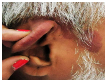
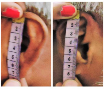
On examination a macular deeply erythematous patch about 8*5 cm in size was seen over the right ear extending to the retro auricular region and the malar area with irregular margins [Figure 1]. No nodules were seen over the lesion. On measuring the right ear (7.5cm length) was larger than the left ear (7cm length) [Figure 2]. Based on these features a diagnosis of port wine stain with megalopinna was made.
3. Discussion
The most common hypothesis for the pathogenesis of port wine stain is the abnormal neural regulation of blood vessels[3]. The may be due to a defect in the embryological maturation of sympathetic fibers leading to a loss of sympathetic control of cutaneous vessels which results in ectasia.
Acquired PWS may occur as a result of herpes zoster infection, tumours, trauma, drugs and sun exposure[4]. This in turn may be due to a loss of sympathetic regulation of cutaneous blood flow. Klippel-Trenaunay-Weber syndrome is a congenital disorder characterized by venous malformation, skeletal tissue hypertrophy and PWS. Gingival hyperplasia has also been associated with PWS in a case of Sturge-Weber syndrome[5]. This syndrome usually presents with PWS over the face, ocular defects, glaucoma and leptomeningeal angioma.
Thus, the megalopinna may be due to vascular ectasia leading to hypertrophy and thus unilateral increase in size of the affected pinna.
4. Conclusion
Ectasia of the dermal blood vessels located in the dermal vascular plexus could lead to tissue hypertrophy and thus megalopinna in cases of port-wine stains located over the pinna.
Treatment of PWS in the early stages would prevent local tissue hypertrophy and thus megalopinna.
References
- Karvnnen SL, Vaajalahti P, Marenk M. Birthmarks in 4346 Finnish newborns. Acta Derm Venereol 1992; 72:55-7
- Mahakrishnan A. Megalopinna in naevus unius lateris: A case report. Acta Derm Venereol 1981; 61(4): 365-7
- Smoiler BR, Rosen S. Port-wine stains. A disease of altered neural modulation of blood vessels? Arch Dermatol 1986; 122:177-9.
- Bansal S, Garg VK, Wadhwa B, Khurana N. Acquired port-wine stain in an adult male: First reported case from India with review of literature. Indian J Dermatol 2015; 60:104
- Reddy PV, Rani KR, Indumathy P, Rajashree D. Gingival overgrowth associated with port-wine stains: A case report of Sturge-Weber syndrome. J NTR Univ Health Sci 2014; 3:287-90.