


|
|
DOI Prefix 10.20431 |
Information
Journal Policies
Biological Post: A Novel Technique to Reinforce a Tooth with Wide Canal Configuration
Dr. Abhipsha Lahiri1*, Dr. Sheetal Ghivari2, Dr. Madhu Pujar3, Dr. Veerendra M. Uppin4
2.Reader, Department of conservative dentistry and endodontics, Maratha Mandal’s Nathajirao G. Halgekar Institute of Dental Science and Research Center, Belagavi, India.
3.Professor and Head, Department of conservative dentistry and endodontics, Maratha Mandal’s Nathajirao G. Halgekar Institute of Dental Science and Research Center, Belagavi, India.
4.Professor, Department of conservative dentistry and endodontics, Maratha Mandal’s Nathajirao G. Halgekar Institute of Dental Science and Research Center, Belagavi, India.
Copyright : © 2018 Authors. This is an open-access article distributed under the terms of the Creative Commons Attribution License, which permits unrestricted use, distribution, and reproduction in any medium, provided the original author and source are credited.
To restore grossly destructed teeth, posts are required to retain the core. Though a variety of materials are available in the market, none of them are ideal. In this case report, a novel post system, i.e. biological post, have been described which was used to reinforce the large canal of a maxillary right central incisor by creating an ideal monoblock followed by core build up and crown placement. This alternative technique offers a feasible low cost option.
Keywords: Biological post, Dentine post, Grossly destructed teeth, Wide canal configuration,Dental Science
1. Introduction
Restoration of grossly destructed endodontically treated anterior teeth is still a challenge. In these teeth post are needed to retain the core material. A material that is used for fabrication of post plays a pivotal role in determining the long term prognosis of the teeth.
Ideally the post material should have physical properties such as modulus of elasticity, thermal expansion and aesthetics that are close to dentin and additionally should bond very well with the dentine [1].
In order to achieve an intraradicular retention various post systems can be used such as custom made cast post or prefabricated posts such as fibre glass, carbon fibre, metal and ceramics.
However, the moduli of elasticity of these posts were 4-7 times higher than that of the dentine and hence showed higher failure rate in anterior teeth [2]. To overcome the disadvantages of these materials, a solution was given in the form of biological post i.e. posts fabricated from dentine.
This case report describes the use of dentine post made from natural extracted sterilized tooth for management of a compromised anterior tooth.
2. Case Report
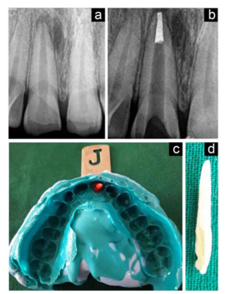
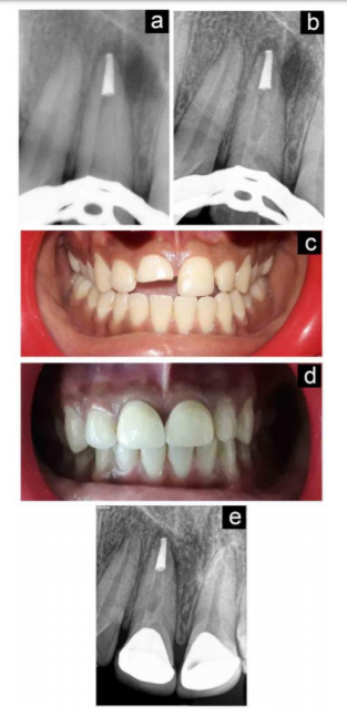
A 19 year old male patient presented with fractured left and right maxillary central incisors upto the junction of cervical and middle third due to trauma one year ago [Figure 2c]. The patient had no relevant medical history. The radiographic examination showed the fracture extending till the pulp space (along with periapical radiolucency) with 11 and involving dentine with 21 [Figure 1a].
Treatment plan proposed to the patient was root canal treatment of 11 followed by placement of post and crown placement of 11 and 21. Placement of fibre post was tried, however the canal was too wide for adaptation of the fibre post. Based on the remaining tooth structure, cast post was also difficult to fabricate, and hence biological post was selected.
Consent of the patient was received to place a post made out of natural extracted human tooth which will be sterilized according to biosecurity standards.
Root canal treatment of 11 was carried out, following which gutta percha was removed from the canal using a peeso reamer till size 4, leaving only 5 mm gutta percha apically [Figure 1b].
Impression of the canal was made with pattern resin (Pattern resin LS, GC, India) and while placing the pattern in the root canal, two step putty and light body silicone impression was made [Figure 1c]. The cast obtained had a replica of the post space prepared in the patient’s tooth.
3. Dentine Post Preparation
An extracted central incisor autoclaved at 121° C for 15 mins at 15lbs psi, was selected for post fabrication. The tooth was sectioned buccolingually using a diamond disc. The previously obtained mould in the master cast was used as reference to orient the shape, thickness, and length of the dentine post [Figure 1d]. The post was then placed in the patient and radiograph was taken to confirm its proper adaptation to the canal space [Figure 2a].
4. Post Cementation
The canal space was cleaned with saline and dried with paper point. Both the canal space and dentine post were coated with a sixth generation bonding agent, Clearfil SE Bond (Kuraray, Japan) which was used according to manufacturer’s instructions.
The luting was done using a self cure resin based luting agent Rely-X U200 self adhesive resin cement (3M ESPE); both the canal space and the post surface were coated with cement. Another radiograph was taken to confirm the proper placement of the post in the canal [Figure 2b]. The core build up was done using a dual cure resin material, FluoroCore 2+ (Densply,Sirona).
5. Tooth Preparation
Tooth preparation was done for porcelain facing metal in both 11 and 21. Impression was recorded using a two step technique using putty and light body silicone based impression material.
6. Crown Cementation
After checking for proper fit and shade match, cementation of the crowns were done using luting type glass ionomer cement (GC Gold Label 1 Luting and lining) [Figure 2d].
Follow up radiograph was taken after one year to confirm proper healing and satisfactory results. [Figure 2e]
7. Discussion
This case report presents intracanal rehabilitation of wide canal configuration in fractured maxillary anterior teeth using a biological post such as dentine post followed by crown cementation. There are various methods to take intracanal impressions to fabricate the biological post such as retrieval of acrylic resin pattern from the root canal of the patient, which is used as a reference to contour the dentine post or direct wax impression of the canal can be used as a guide for shaping the dentine post. In present case report we have adopted the first method as reported by Faria et al [3].
The extracted tooth for preparation of dentine post was selected from patients scheduled for extraction of intact maxillary central incisor due to periodontal disease. After thorough medical history of the donor, routine blood investigations for systemic infections such as Human immunodeficiency virus, Hepatitis B virus were done. Following extraction the tooth was properly cleaned and autoclaved at 121°C under 15lbs psi for 15 minutes ensuring all biosecurity standards. As per the various studies, biomechanical properties of dentine in freshly extracted teeth were preserved although autoclaving may cause slight variation in dentine micro hardness[4,5].
The physical properties of dentine post closely resemble root dentine for their various properties such as modulus of elasticity; viscoelastic and thermal properties, compressive strength. According to studies conducted by Ambica et al[6] and Kathuria et al [7] fracture toughness of dentine was found to be better then various recent restorative materials. Dentine post forms a uniform monoblock which helps in even distribution of stress across the root dentine [8,9]. Due to similarity in the elasticity of dentine post to that of root dentine, it allows the post flexion to mimic tooth flexion which minimises the stress concentration and root fracture [10]. As this biological post system is less expensive it may be best alternative to the people of lower economic strata.
8. Declaration Of Patient Consent
The authors certify that they have obtained all appropriate patient consent forms. In the form the patient(s) has/have given his/her/their consent for his/her/their images and other clinical information to be reported in the journal. The patients understand that their names and initials will not be published and due efforts will be made to conceal their identity, but anonymity cannot be guaranteed.
9. Conclusion
This case report has described a case where a post made out of human dentine was used to reinforce a fractured anterior tooth in order to restore its form, function and esthetics. It is a novel and economic treatment option to restore grossly mutilated anterior teeth.
References
- Cheung W. A review of the management of endodontically treated teeth: post, core and the final restoration. J Am Dent Assoc 2005;136:611–9.
- Newman MP, Yaman P, Dennison J, et al. Fracture resistance of endodontically treated teeth restored with composite posts. J Prosthet Dent 2003;89:360–7.
- Correa-Faria P, Alcantara CE, Caldas-Diniz MV, Botelho AM, Tavano KT. “Biological Restoration”: Root Canal and Coronal Reconstruction. J Esthet Restor Dent. 2010;22(3):168-77.
- Pantera EA, Schuster GS. Sterilization of extracted human teeth. J Dent Educ. 1990;54:283–5.
- Parsell DE, Stewart BM, Barker JR, Nick TG, Karns L, Johnson RB. The effect of steam sterilization on the physical properties and perceived cutting characteristics of extracted teeth. J Dent Educ. 1998;62:260–3.
- Ambica K, Mabendran K, Talwar S, Verma M, Padmini g, Periasamy R. Static and fatigue loading of endodontically treated teeth restored with carbon fiber posts, glass fiber posts, and an experimental dentin post system: An in vitro study. J Endod. 2013; 39(1):96-100 7.
- Kathuria A, Kavitha M, Khetarpal S. Ex vivo fracture resistance of endodontically treated maxillary central incisors restored with fiber-reinforced composite posts and experimental dentin posts. J Conserv Dent. 2011; 14(4):401-05.
- Kaizer OB, Bonfante G, Pereira Filho LD, et al. Utilization of biological posts to reconstruct weakened roots. Rev Gaucha Odontol 2008; 56:7–13.
- Batista A, Lopes CG. Performed dentin post reinforcing teeth with immature apexes. Rev Bras Prot Clin Lab 1999;3:199–21.
- Martelli R. Fourth-generation intraradicular posts for the aesthetic restoration of anterior teeth. Pract Periodontics Aesthet Dent 2000;12:579-84.