


|
|
DOI Prefix 10.20431 |
Information
Journal Policies
"A New Technique in Model Planning for Orthognathic Surgery"
Fatih Celebi1*, Sibel Akbulut1
Copyright : © 2018 Authors. This is an open-access article distributed under the terms of the Creative Commons Attribution License, which permits unrestricted use, distribution, and reproduction in any medium, provided the original author and source are credited.
Objective: The aim of this study is to introduce a new technique that can improve the reliability of the model planning for orthognathic surgery.
Materials and Methods: A semi-adjustable articulator, a pocket-size digital inclinometer, and a laser alignment device are required for this method. Presurgical records, including extraoral and intraoral photos, radiographs, alginate impressions, and centric bite registration were acquired at latest 15 days prior to the surgery. Cephalometric planning is carried out. The position of the maxilla, according to the axis-orbital plane, is registered through the facebow and transferred to the semi-adjustable articulator. Vertical and horizontal incision lines are registered on the mounted models by using the laser alignment device.
Results: Using the technique presented in this article, it is possible to transfer the genuine true horizontal and vertical planes to articulator and surgical models.
Conclusion: We assert that this method will reduce the inconsistency between the expected and the actual surgical outcomes.
Keywords: Model planning; Orthognathic surgery ,Dental Science
1. Introduction
Accuracy in planning leads to successful outcomes in treatment. This assumption is particularly important in orthognathic surgery, as the stress of the operation and the lack of consciousness of the patient while under general anesthesia mean that it is impossible to evaluate the results and modify the surgery. Therefore, accurate preoperative records, and accurate planning created by those records, play a key role in orthognathic surgery.
The amount of movement in different directions is currently determined using computerized prediction software, after which intermediate and final splints are constructed with the aid of model surgery; a semi-adjustable articulator is used to construct the intermediate splint. The face bow apparatus, which is a part of the semi-adjustable articulator, is utilized for registering the position of the maxilla according to the skull (the axis-orbital plane)[1,2] and transferring this position to the articulator.
Planning for bimaxillary surgery is more complicated and more sensitive to planning errors, compared with planning for single-jaw surgery. In the latter, only one splint is required, and it can be fabricated using even a non-adjustable articulator. However, in bimaxillary surgery, two splints (intermediate and final) are required, and the intermediate splint cannot be fabricated without using the semi-adjustable articulator[3]; while the occlusal plane of the jaw that is not undergoing surgery can serve as the reference plane for the movement of the jaw being operated on in single-jaw surgery, a usable reference plane is absent in bimaxillary jaw surgery. Therefore, a preoperative occlusal plane relative to the areas not planned for surgery (the axis-orbital plane) must be registered, and a semi-adjustable articulator that can perform this task, must be used.
It has previously been stated that there could be an inconsistency between the planned and the occurring jaw movements in orthognathic surgery, and errors in facebow transfer and model surgery have primarily been cited as the main reasons for this inaccuracy[4]. The present study introduces a new method for utilization in bimaxillary jaw surgery, and we assert that it will increase the credibility of model surgery.
2. Application Of The Method
A semi-adjustable articulator, a pocket-size digital inclinometer, and a laser alignment device are required for this method. Presurgical records, including extraoral and intraoral photos, radiographs, alginate impressions, and centric bite registration were acquired at latest 15 days prior to the surgery. Cephalometric planning is manually or digitally carried out. The position of the maxilla, according to the axis-orbital plane, is registered through the facebow and transferred to the semi-adjustable articulator. Plaster models, which are acquired from alginate impressions, are mounted on the articulator with the guidance of the facebow and centric bite registration. The part of the procedure described up to now is standard, and is carried out in this manner, with minor modifications, in almost every clinic.
The incision line on the mounted models is critical at this point. Some clinicians prefer to take the upper part of the articulator as a guide for this line, while others assert that the occlusal plane must be taken as a guide, as a Le Fort 1 incision is almost always carried out according to the occlusal plane5, and inconsistency between the model incision line and the actual surgery line can result in mismatching operated segments.
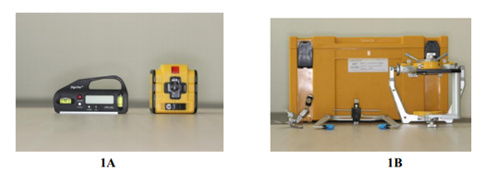
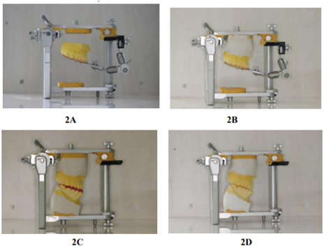
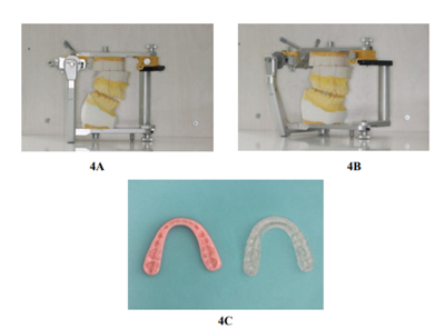
In this article, we suggest a new guide that can identify true horizontal and vertical lines on the mounted models. First, the articulator is placed on a platform. A pocket-size digital inclinometer is then used to ensure that this platform has no inclination, and the required minor changes are made to the platform until the inclinometer indicates 0°. The laser alignment device is then started, and the true horizontal and vertical lines are marked using the marker pen. The maxillary plaster model is separated from the articulator and the model is cut off at the marked line. Vaseline is applied to the upper part of the model that has been prepared by cutting, and it is again mounted on the articulator. The plaster is poured into the space that has been created as a result of the cut off between the upper part of the model and the articulator. Vaselination allows the true horizontal line, registered by the laser alignment device, to serve as the incision line on the model. Consequently, the desired vertical and sagittal movements can be reliably achieved using the reference lines, and the proper intermediate splint can be prepared.
3. Case Report
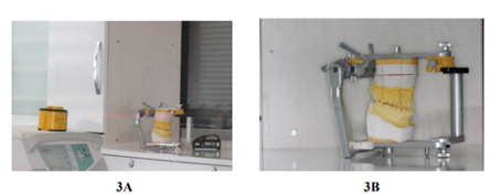
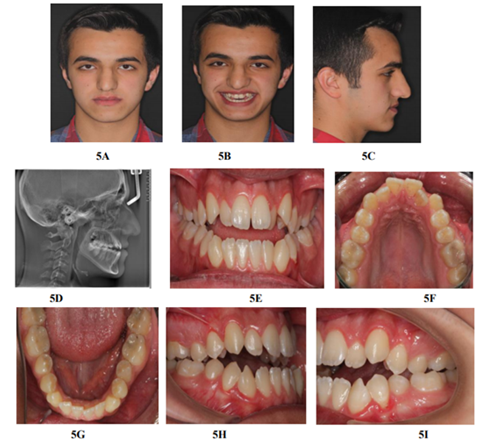
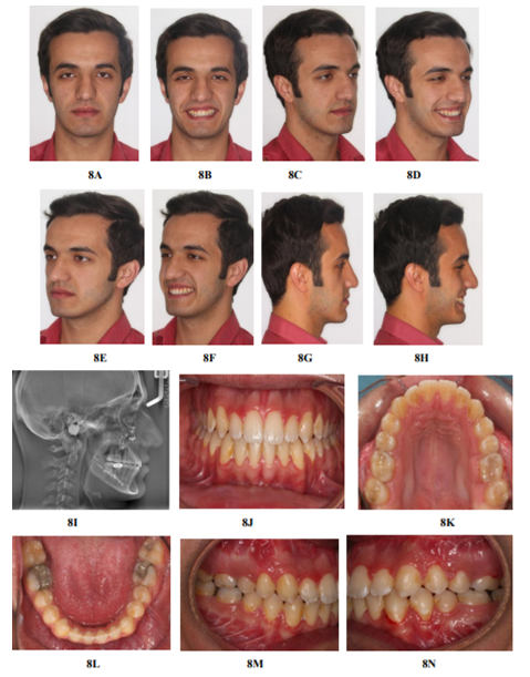
A 16-year-old male presented at our clinic with a chief complaint of anterior open bite. He had a straight profile, insufficient maxillary projection, an under-projected nasal tip, an acute nasolabial angle, competent lips, backward rotation of the mandible and increased lower face height. A class III molar relationship, anterior open bite, posterior cross bite, and mild dental arch crowding were found during intraoral examination. A radiographic data set is presented in Table 1. A combination of orthodontic treatment and orthognathic surgery was the planned approach.
4. Treatment Progress
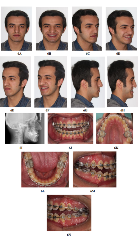
An .022" X .028" Roth-prescription preadjusted fixed appliance was bonded; .012", .14", .16", .017" × .025" nickel-titanium and .017" × .025" stainless steel archwires served in the levelling and aligning stage, and the upper and lower third molars were extracted. In the intermediate stage of the fixed treatment, the transpalatal arch was placed for obtaining the buccal root torque.
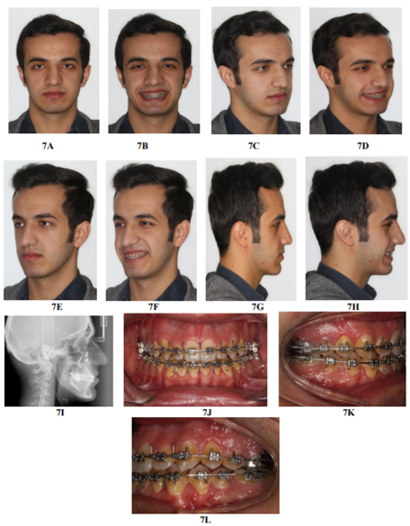
When we thought that surgery was all set to go ahead, the patient stated that he wanted to postpone his operation until the semester holiday, so we had to delay surgery for 5 months. Presurgical records, including extraoral and intraoral photos, radiographs, alginate impressions, and centric bite registration, were acquired on the 15th day before the semester holiday. The evaluation of these records allowed us to carry out final surgical planning: double-jaw surgery, which could achieve a 7 mm maxillary advancement and a counter clockwise mandibular rotation. Intermediate and final splints were manufactured. The intermediate splint was prepared as described above. Upper and lower .018" X .025" stainless steel archwires were inserted and crimpable hooks were placed where required. Surgery was performed under general anesthesia. The final splint was used after the surgery for a while to stabilize the jaws. The fixed appliance was consequently removed, and lingual retainers were placed in both the upper and lower dental arches. Total treatment time was 37 months.
5. Discussion
In terms of perfection in reaching surgical goals, the direction of the incision line in surgery is important, but the direction of the incision line in model surgery has greater significance. In the worst-case scenario, an error in the incision line in surgery leads to the mismatch of the segments on which the operation has been carried out and it does not result in problems in obtaining the planned movement; a well-prepared splint guides the surgeon, such that they achieve accurate jaw movement. However, an error in the incision line in model surgery leads to an improperly prepared splint and unsatisfactory results[3].
The maxilla and the occlusal plane rotate clockwise relative to the true horizontal plane in the majority of orthognathic patients. Therefore, if the occlusal plane or apexes of the teeth serve as guidance in carrying out the incision in model surgery, sagittal movements (advancement or set back) will also result in vertical movements. When the presurgical status of the case described in the present article is examined, it must be noted that the patient was at risk of a gummy smile as a result of maxillary advancement surgery. He already had a mild gummy smile prior to surgery, and if we had performed a downward movement in addition to advancement, a moderate or severe gummy smile would have been inevitable. When examining the final records, it can be observed that this did not occur. Using our new technique, the incision line was positioned parallel to the true horizontal plane, which had been registered using the facebow. Therefore, if the axis-orbital plane is accepted as the true horizontal plane, pure sagittal movement can be achieved without unplanned vertical movements.
Using the technique presented in this article, it is possible to transfer the genuine true horizontal and vertical planes to articulator and surgical models. We assert that this method will reduce the inconsistency between the expected and the actual surgical outcomes. With minor modifications in clinical practice, it can also be useful for a variety of different aims in which identification of the true horizontal and vertical planes is required.
References
- Ellis E, Tharanon W, Gambrell K. Accuracy of face bow transfer: Effects on surgical prediction and post surgical result. J Oral Maxillofac Surg 1992: 50: 562–567.
- Gateno J, Forrest KK, Camp B. A comparison of three methods of face bow transfer recording: Implication for orthognathic surgery. J Oral Maxillofac Surg 2001: 59: 635–640.
- Hammoudeh JA, Howell LK, Boutros S, Scott MA, Urata MM. Current Status of Surgical Planning for Orthognathic Surgery: Traditional Methods versus 3D Surgical Planning. Plast Reconstr Surg Glob Open. 2015 Mar 6;3(2):e307.
- Sharifi A, Jones R, Ayoub A, Moos K, Walker F, Khambay B, McHugh S. How accurate is model planning for orthognathic surgery? Int J Oral Maxillofac Surg. 2008 Dec;37(12):1089-93.
- Ehmer U, Joos U, Ziebura T, Flieger S, Wiech mann D. The University Münster Model Surgery System for Orthognathic Surgery. Part II -- KD-MMS. Head Face Med. 2013 Jan 4;9:2.