


|
|
DOI Prefix 10.20431 |
Information
Journal Policies
Left Atrial Appendage Absence: Multimodality Evaluation
A.Cecchetto1*, C. Ciccio2, G. Molon1, A. Costa1, S. Bonapace1, G. Carbognin2, E. Barbieri1, L. Lanzoni1
2.Radiology Dept, SacroCuore Don Calabria Hospital, Negrar, Verona, Italy.
Copyright : © 2018 Authors. This is an open-access article distributed under the terms of the Creative Commons Attribution License, which permits unrestricted use, distribution, and reproduction in any medium, provided the original author and source are credited.
Among possible anatomical variants, agenesia of the left atrial appendage (LAA) is very rare and differential diagnosis for no visualization of the LAA during transesophageal echocardiogram (TEE) includes complete occlusion by thrombus, poor echo cardiographic windows, prior surgical ligation or percutaneous closure. We presented a case of a patient with absence of left atrial appendage evaluated through multiple imaging techniques.
Appendage Absence, Multimodality Evaluation,Cardiology
Clinical Case
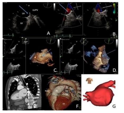
A 69 years old man, with persistent atrial fibrillation (AF) was referred to our Centre for cryoablation procedure. His work up included two dimensional (2D)/ three dimensional (3D) TEEto exclude intracardiac thrombus and contrast-enhanced multidetector computed tomography (CT) scan with Navx navigation system of reconstruction for anatomical details of the left atrium (LA). Despite 2D TEE imaging at multiple angles, LAA could not be visualized (arrows, Figures 1A) and colour Doppler imaging characteristic flow of LAA could not be demonstrated (Figure 1B). 3D TEE "en face" view demonstrated the absence of LAA anatomic orifice, below left upper pulmonary vein and lateral ridge with uniformity appearance of LA lateral wall (Figure 1C). Cropping 3D data sets, no LAA body was identified, only a small residue (Figure 1D). Multidetector CT scan confirmed the suspicion of LAA absence with optimal spatial resolution (Figures 1E, 1F). Anatomical reconstruction of LA during cryoablation procedure led to the same conclusion, showing pulmonary veins in the usual anatomic pattern and LAA body absence on LA outer surface (Figures1G).
LAA contributes toward left atrial reservoir and contractile functions. However, LAA is also the most common site for thrombus formation in AF. The shape of the LAA is variable, but the absence is extremely rare [1-3]. 3D TEE and multidetector CT could be performed to confirm the presence or absence of LAA and to exclude thrombotic occlusion especially in the era of LA intervention [4,5]. Physiological consequences and the impact on cardio-embolic risk of a congenitally absent LAA are unknown and it seems more likely congenital anatomical variation.
References
- Collier P, Cavalcante JL, Phelan D, et al. Congenital absence of the left atrial appendage. CircCardiovasc Imaging 2012; 5:549-50.
- Zhang ZJ, Dong JZ, Ma CS. Congenital absence of the left atrial appendage: a rare anatomical variation with clinical significance. Acta Cardiol 2013; 68:325-7.
- Di Gioia G, Mega S, Visconti S, et al. Congenital absence of left atrial appendage in a patient with intracranial hemorrhage. Am J Case Rep 2015; 16: 514-6.
- Saleh M, Balakrishnan R, Kontak LC, et al. Congenital absence of the left atrial appendage visualized by 3D echocardiography in two adult patients. Echocardiography 2015; 32: 1206-10.
- Di Biase L, Burkhardt JD, Mohanty P, et al. Left atrial appendage isolation in patients with longstanding persistent AF undergoing catheter ablation: BELIEF trial. J Am CollCardiol 2016; 68: 1929-40
- Song IG, Kim SH, Oh YS, Rho TH. Underdevelopment of left atrial appendage. Korean Circ J 2017; 47:141-3.