


|
|
DOI Prefix 10.20431 |
Information
Journal Policies
ARC Journal of Anesthesiology
Volume-2 Issue-2, 2017, Page No: 5-6
Advik Technique' of Peripheral Intravenous Cannulation-Different Perspective in Difficult Situations
Bhavna Gupta1,Kamna kakkar2,Anish Gupta3,Lalit Gupta4
1.Senior resident, deptt of anaesthesia, MAMC AND LOK NAYAK HOSPITAL.
2.PG student, deptt of anesthesia PGIMS ROHTAK.
3.Senior resident CTVS, AIIMS.
4.Assistant professor, dept of anaesthesia, MAMC AND LOK NAYAK HOSPITAL.
2.PG student, deptt of anesthesia PGIMS ROHTAK.
3.Senior resident CTVS, AIIMS.
4.Assistant professor, dept of anaesthesia, MAMC AND LOK NAYAK HOSPITAL.
Citation : Bhavna Gupta,Kamna kakkar,Anish Gupta,Lalit Gupta, "Advik Technique’ of Peripheral Intravenous Cannulation-Different Perspective in Difficult Situations" ARC Journal of Anesthesiology. 2017; 2(2):5-6.
Copyright : © 2017 Authors. This is an open-access article distributed under the terms of the Creative Commons Attribution License, which permits unrestricted use, distribution, and reproduction in any medium, provided the original author and source are credited.
Advik Technique,Peripheral Intravenous,Cannulation-Different Perspective,Anesthesiology
Editorial
Peripheral intravenous cannulation is an essential part of management of patients in hospitals, especially in emergency-department. Steps of intravenous cannulation includes: sanitization of operator’s hands, followed by tourniquet placement, identification of vein by placing the extremity in dependent position, and asking the patient to make a tight fist. Skin of the patient is prepped with antiseptic solution. After the vein becomes prominent, needle of cannula is inserted with bevel facing upwards until a flashback of blood is seen in the hub of cannula. Once this is seen, the cannula is progressed, needle is fixed while the rest of the cannula is advanced gently into the vein.
Peripheral venous cannulation is basic but critical component of patient care. It a simple, reliable, and inexpensive method to save a patient’s life in need. It is an art which is learnt with practice and experience. Even in experienced hands, it may fail in difficult patient subsets. The common difficulties encountered in establishing the intravenous-access include movement of patient during cannulation, presence of venous valves obscuring passage of catheter, short or tortuous course of vein, etc. In case of failed cannulation, it is a traditional practice to remove entire cannula and start again. It leads to bleeding at the punctured site. Moreover, tourniquet in situ leads to further bleeding, and consequently it is released. More time is misspent to apply pressure on the punctured site using a sterile gauze-piece to stop the bleed. After losing precious time in securing hemostasis in the previously punctured site, a second attempt is made to cannulate the patient. Tourniquet is again applied at a separate site and, the entire process is repeated again. This practice is time consuming and usually, requires help of assistant to reduce the overall time taken to achieve successful cannulation.
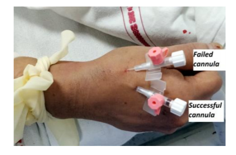
We deduced a novel ‘Advik technique’ of peripheral intravenous cannulation in difficult or failed situations. In our technique, when we encounter a failed cannula situation, we leave the catheter of the cannula in situ, remove its needle and cap the catheter. There is no need to release the tourniquet or apply pressure on the punctured site, as the capped cannula itself helps to achieve hemostasis of its punctured site. In the meanwhile, with the tourniquet still in place, we find another vein- adjacent or nearby and insert a second cannula in it. [Figure 1] We fill a syringe with saline and flush it through the injection port of this second cannula to check its patency and ensure successful cannulation. After successfully inserting the second cannula, tourniquet is removed. After this, the previously failed cannula is now removed. Sterile gauze is pressed over the punctured site and hemostasis is achieved.
Peripheral intravenous cannulation is an essential part of management of patients in hospitals, especially in emergency-department. Steps of intravenous cannulation includes: sanitization of operator’s hands, followed by tourniquet placement, identification of vein by placing the extremity in dependent position, and asking the patient to make a tight fist. Skin of the patient is prepped with antiseptic solution. After the vein becomes prominent, needle of cannula is inserted with bevel facing upwards until a flashback of blood is seen in the hub of cannula. Once this is seen, the cannula is progressed, needle is fixed while the rest of the cannula is advanced gently into the vein.
Peripheral venous cannulation is basic but critical component of patient care. It a simple, reliable, and inexpensive method to save a patient’s life in need. It is an art which is learnt with practice and experience. Even in experienced hands, it may fail in difficult patient subsets. The common difficulties encountered in establishing the intravenous-access include movement of patient during cannulation, presence of venous valves obscuring passage of catheter, short or tortuous course of vein, etc. In case of failed cannulation, it is a traditional practice to remove entire cannula and start again. It leads to bleeding at the punctured site. Moreover, tourniquet in situ leads to further bleeding, and consequently it is released. More time is misspent to apply pressure on the punctured site using a sterile gauze-piece to stop the bleed. After losing precious time in securing hemostasis in the previously punctured site, a second attempt is made to cannulate the patient. Tourniquet is again applied at a separate site and, the entire process is repeated again. This practice is time consuming and usually, requires help of assistant to reduce the overall time taken to achieve successful cannulation.
We deduced a novel ‘Advik technique’ of peripheral intravenous cannulation in difficult or failed situations. In our technique, when we encounter a failed cannula situation, we leave the catheter of the cannula in situ, remove its needle and cap the catheter. There is no need to release the tourniquet or apply pressure on the punctured site, as the capped cannula itself helps to achieve hemostasis of its punctured site. In the meanwhile, with the tourniquet still in place, we find another vein- adjacent or nearby and insert a second cannula in it. [Figure 1] We fill a syringe with saline and flush it through the injection port of this second cannula to check its patency and ensure successful cannulation. After successfully inserting the second cannula, tourniquet is removed. After this, the previously failed cannula is now removed. Sterile gauze is pressed over the punctured site and hemostasis is achieved.
Figure1