


|
|
DOI Prefix 10.20431 |
Information
Journal Policies
ARC Journal of Cancer Science
Volume-2 Issue-1, 2016
Abstract
Oral cancer is most common cancer in males and third most common in females, the main causative agent being use of chewing betel quid (BQ). The micronucleus (MN) assay in exfoliated buccal cells is a useful and minimally invasive method for monitoring genetic damage in humans. Micronuclei (MN) have been proposed as good biomarker to assess cytogenetic damage. MN formation has been observed in cancer and pre-cancerous lesions of the oral cavity of betel quid chewers. In this present study cases were screened from Camp in Eastern India, Camp in North East India, Subjects attending Oral &Maxillofacial and ENT department of Ramakrishna Mission Seva Pratishthan(RKMSP), Kolkata and Onco surgery department, ESI hospital, Sealdah, Kolkata. Micronuclei percentage are higher in oral cancer cases than normal.
2.KEYWORDS
3.INTRODUCTION
4.MATERIALS AND METHODS
5.ORAL SMEAR WERE OBTAINED FROM SUBJECTS AS FOLLOWS
5.1.Parameters for cell inclusion in the cells to be scored
5.2.Parameters for identifying micronucleus
6.RESULTS
7.DISCUSSION
8.ACKNOWLEDGEMENT
9.REFERENCES
AUTHOR DETAILS
Aniket Adhikari*, Madhusnata De.
Department of Genetics, Vivekananda Institute of Medical Sciences,
Ramakrishna Mission Seva Pratishthan. 99, Sarat Bose Road, Kolkata – 700026. India.
*[email protected]
KEYWORDS
Oral cancer, betel quid, micronuclei
INTRODUCTION
The cytogenetic assay of micronuclei was established almost thirty years ago [1]. Micronuclei (MN) are structures that arise from acentric chromo-some fragments or complete chromosomes that failed to attach to mitotic spindle during cytokinesis and are excluded from the daughter nuclei into the cytoplasm [2,4]. MN represent structural chromosomal aberrations (chromosome loss or breakage) induced by ionizing radiation or chemical mutagens [3]. Micronuclei can be measured after DNA staining by the Feulgen reaction, Giemsa or fluore-scence dyes.
At present, the micronucleui test is the most popular short-term assay for evaluation of clastogenicity [5,-9]. Micronuclei arise from chromosomal fragments or chromosomes that are not incorporated into daughter nuclei at the time of cell division.DNA damage can be assessed by cytogenetic markers like chromosomal aberrations, sister chromatid exchanges and micronuclei. Out of all these, micronucleus test is preferable as it does not require tedious procedures like cell culture and metaphase preparation. To further add, as it is applicable on interphase cells only, it is the best indicator of mitotic interference and chromosomal mutations or breakages. It is a non-invasive and very economical procedure [10].
In the early 1970s, the term micronucleus was first time suggested by Boller and Schmidt and Heddle who showed that this is a simple method to detect genotoxic potential of mutagens after in vivo exposure of animals using bone marrow erythrocytes [11]. About 25 years ago, Stich and co-workers developed a protocol for micronucleus assay with exfoliated human epithelial cells which has been widely used in occupational & lifestyle studies [12].The buccal cell micronucleus assay was proposed in 1983, thereafter it gained popularity as a biomarker of genetic damage in numerous applications. A few years later, Countryman & Heddle showed that peripheral blood lymphocytes could be used for micronucleus approach and recommended to use micronuclei as a biomarker in testing schemes. Theodor Boveri originally observed the fact that abnormal nuclear morphologies commonly occur in cancer. Micronuclei are also referred to Howell-Jolly bodies; discovered by hematologists William Henry Howell and Justin Marie Jolly in erythrocytes. Micronucleus induction by a chemical was first reported in Ehrlich ascites tumor cells treated with colchicine. In anaphase, the microtubules are not attached properly to the chromosomes, which can cause pulling in a different direction. This results in parts of the chromatids or chromosomes being broken off and enveloped as an extra nucleus in one of the daughter cells. This is the main way that micronuclei are formed. Cancer, modern epidemic among non-communicable diseases is the second commonest cause of mortality in developed countries and remains one of the ten commonest causes of mortality in developing countries like India, is a complex disease with altered expression, abnormal growth and disruption of normal function of cells caused by genotoxic effects of chemical carcinogens or environmental pollutants resulting in genomic instability at an early stage of cancer, which is reflected often as leukoplakia, erythroplakia, Lichen planus and sub mucous fibrosis. Oral squamous cell carcinoma and the most common oral pre malignancies such as leukoplakia and oral submucous fibrosis appear to be related to the habit of betel quid (BQ) chewing in South East Asia, whereas in Western countries cigarette smoking and heavy alcohol consumption are the main causative agents. Areca nut (Areca catechu), a major component of BQ. Areca nut contains certain alkaloids that give rise to nitrosamines, some of which such as N nitrosoguvacoline, 3-(methylnitrosamino) propionitrile, 3-methylnitrosaminopropionaldehyde and N-nitrosoguvacine, are shown to be carcinogenic [16]. These BQ-specific nitrosamines may act as an adjunct to tobacco specific nitrosamines that are strongly implicated as an etiologic factor for leukoplakia and oral submucous fibrosis.
MATERIALS AND METHODS
Screening of subjects was carried out from Camp in Eastern India, Camp in North East India,Subjects attending Maxillofacial and ENT department of Ramakrishna Mission Seva Pratishthan(RKMSP), Kolkata and Onco surgery department, ESI hospital, Sealdah, Kolkata.
An informed consent were taken from all the subjects and approval by Institute Ethical Committee.
More than 2000 cases were screened from various camps and hospitals (RKMSP and ESI). 92 cases had betel quid addiction with cancerous and pre cancerous cases (with lesion) from various camps and hospitals and 22 cases had no betel quid addiction as control.
ORAL SMEAR WERE OBTAINED FROM SUBJECTS AS FOLLOWS
There are several factors that affect the MN in exfoliated oral mucosal cells like differences in timing and implements used in cell collection, fixation, staining techniques, number of cells counted, scoring criteria, and other nuclear abnormalities in normal or degenerated cells [14].
To obtain a smear from the oral cavity pre moistened wooden spatula can be used. It is also advised to rinse the oral cavity before collection of the samples to remove the food debris and necrotic slough, if any, which could alter the quality of the smear [15].
Once the exfoliated cells are collected, they are smeared immediately on pre-cleaned microscopic slides followed by fixation in 80% methanol [11] .Tolbert et al, also recommended the scoring of at least 1000 cells, which can be increased to 2000-3000 if less than 5 micro nucleated (MN) cells are observed after counting 1000 cells6 to determine the MN Percentage.
In 1992 Tolbert et al developed the criteria for choosing the cells which consists of the following parameters [13].
i. Intact cytoplasm and relatively flat cell position on the slide.
ii. Little or no overlap with adjacent cells.
iii. Little or no debris.
iv. Nucleus normal and intact, nuclear perimeter smooth and distinct.
i. Rounded smooth perimeter suggestive of a membrane.
ii. Less than a third the diameter of associated nucleus, but large enough to discern shape and color.
iii. Staining intensity similar to nucleus.
iv. Texture similar to nucleus.
v. Same focal plane as nucleus.
vi. Absence of overlap with bridge to nucleus.
RESULTS
Inference: Micronuclei (MN) which acts as a cancer biomarker, 5 folds increase than normal in cancer cases who had betel quid chewing habit.
DISCUSSION
Micronuclei assay is a sensitive, non-invasive and low cost technique that offers a very simple method for obtaining information on status of the epithelial cells, particularly DNA damage, proliferative potential of basal cells and cell death. Although many studies have shown a statistically significant increased MN frequency in the buccal cells of populations exposed to occupational and environmental insults, various lifestyle factors and oral cancer. MN, which acts as a cancer biomarker, highly increase than normal in cancer cases and pre cancerous cases who had betel quid chewing habit.
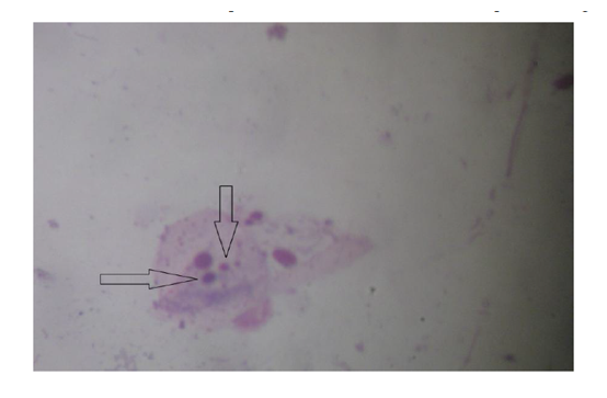
Figure 1. Buccal Smear Showing Micronuclei (MN)
ACKNOWLEDGEMENT
The authors thank the Secretary, Ramakrishna Mission Seva Pratishthan, for kind permission to use the laboratory for this work and Department of Maxillofacial and ENT Department of RKMSP Hospital and also Dr.P.B.Kar, Dr. A.Das, Dr. Johuri of ESI Hospital, Sealdah ,Kolkata.
REFERENCES
- Schmid W. The micronucleus test. Mutat Res. 31:9-15(1975).
- Weissenborn U, Streffer C .Micronuclei with kinetochores in human melanoma cells and rectal carcinomas. Int J Radiat Biol 59:373-83(1991).
- Fenech M. The in vitro micronucleus technique.Mutat Res 455:81-95 (2000).
- Fuhrmann C, Streffer C, Müller W-U, Becker U (1992).Micronucleus assay prediction and application optimized by cytochalas in B-induced binucleated tumor cells. Strahlenther Onkol 168:603-9.
- Yoshikawa T, Tanigawa M, Tanigawa T, Imai A, Hongo H & Kondo M, Enhancement of nitric oxide generation by low frequency electromagnetic field, Pathophysiology, 7: 131(2000).
- Mc Namee J P, Bellier P V, Gajda G B, Miller S M, Lemay E P, Lavallee B F, Mano L & Thansandote A, DNA damage and micronucleus induction in human leukocytes after acute in vitro exposure to 1.9 GHz continuous wave radiofrequency field, Radiation Research, 158 :523 (2002).
- Kumar S, Kesari K K & Behari J, Evaluation of genotoxic effects in male wistar rats following microwave exposure, Indian J Exp Biol, 48:586(2010).
- Kesari K K, Kumar S & Behari J, Effects of radiofrequency electromagnetic waves exposure from cellular phone on reproductive pattern in male Wistar rats, Appl Biochem Biotechnol, 164:546 (2011).
- Desai N, Kesari K K & Agarwal A, Pathophysiology of cell phone radiation: oxidative stress and carcinogenesis with focus on male reproductive system, Reprod Biol Endocrinol, 7 : 114 (2009).
- Pratheepa S N,Kaur S, Reddy KS, Vivekanandam S, Rao RK. Micronucleus Index: An early diagnosis in oral carcinoma. J Anat Soc India; 57(1):8-13(2008).
- Kashyap B, Reddy PS. Micronuclei assay of exfoliated oral buccal cells: means to assess the nuclear abnormalities in different diseases. J Cancer Res Ther; 8(2):184-191(2012).
- Nersesyan AK, Ilin AI. The micronucleus assay in exfoliated human cells: A mini review of papers from the CIS. Cytology and Genetics.;41(2):115-124(2007).
- Palaskar S, Jindal C. Evaluation of micronuclei using Papanicolaou and May Grunwald Giemsa stain in individuals with different tobacco habits: A comparative study. J Clin DiagRes;4:3607–3613(2010).
- Mahimkar MB, Samant TA, Kannan S , Patil T. Influence of genetic polymorphisms on frequency of micronucleated buccal epithelial cells in Leukoplakia patients. Oral Oncol; 46(10):761–766(2010).
- Jadhav K, Gupta N, Ahmed MB. Micronuclei: An essential biomarker in oral exfoliated cells for grading of oral squamous cell carcinoma. J Cytol; 28(1):7–12(2011).
- Hoffmann D, Brunnemann KD, Prokopczyk B, Djordjevic MV. Tobacco-specific N nitrosamines and Areca-derived Nnitrosamines: chemistry, biochemistry, carcinogenicity, and relevance to humans. J. Toxicol Environ Health. 41:1-52(1994).